Which bone bears the superior and inferior temporal lines
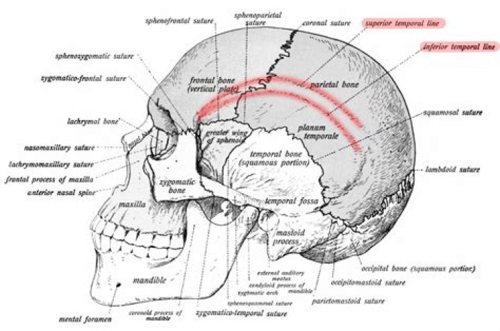
BordersSagittal (superiorly), squamosal (inferiorly), frontal (anteriorly), occipital (posteriorly)Cranial suturesSagittal, sphenoparietal, parietomastoid, coronal, lambdoidExternal surfaceSuperior and inferior temporal lines, parietal eminence, temporal fossa, parietal foramen
Which bone bears the Infraorbital foramen?
In human anatomy, the infraorbital foramen is an opening in the maxillary bone of the skull located below the infraorbital margin of the orbit. It transmits the infraorbital artery and vein, and the infraorbital nerve, a branch of the maxillary nerve.
What cranial and facial bones may be seen easily on the inferior surface of the skull quizlet?
(b) The hard palate, the sphenoid bone, parts of the temporal bone, and the occipital bone with its foramen magnum may be seen in the inferior view.
Which skull bone Cannot palpate?
The skull consists of _____ cranial bones and _____ facial bones.8,14which of these skull bones cannot be palpatedEthmoidwhich of these bones does not contribute to the region known as the pterionOccipitalwhich bones form the calvariaoccipital, parietal, and frontalWhich cranial fossa supports the cerebellum quizlet?
Which cranial fossa supports the cerebellum? middle cranial fossa. The basin in the floor of the cranium that accommodates the temporal lobe of the brain is the: posterior cranial fossa.
What is inferior orbital fissure?
The inferior orbital fissure is formed by the sphenoid bone and the maxilla. It is located posteriorly along the boundary of the floor and lateral wall of the orbit. It transmits a number of structures, including: the zygomatic branch of the maxillary nerve. the ascending branches from the pterygopalatine ganglion.
Which bones form the inferior margin of the orbit?
The inferior orbital fissure lies on the floor of the orbit. The superior border is the greater wing of the sphenoid, and the maxilla and palatine bone compose the inferior border, with the zygomatic bone laterally.
Which bone is not part of the axial skeleton?
The humerus is the bone that makes up the upper arm of both upper extremities. This bone is part of the appendicular skeleton. Therefore, the correct answer is (c) Humerus.What bone bears the mandibular fossa?
In hominids, the mandibular fossa is located on the temporal bone on the lateralmost edge of the skull base. It forms the cranial portion of the temporomandibular joint, and is formed largely by the squamous portion of the temporal bone.
On what bone are the superior nasal Conchae found quizlet?The inferior portion of the sphenoid bone forms the superior nasal concha. The nasal concha are located on the medial wall of the nasal cavity. All of the nasal concha are part of the same bone.
Article first time published onWhich bones are visible while looking at a superior view of the skull?
This view of the skull base demonstrates the occipital, temporal and sphenoid bones. The petrous portion of the temporal bone articulates in the posterior fossa with the occipital bone, and the squamosal portion of the temporal bone articulates with the sphenoid at the sphenosquamosal suture.
Which suture forms the boundary between the temporal bone and the parietal bone of that side?
Squamosal suture: the suture between the temporal and parietal bones.
What is the name of the keystone shaped plane of the forehead that is found between the two eyes?
GlabellaSide view of head, showing surface relations of bones (glabella labeled at center left)DetailsIdentifiersLatinGlabella
What is the correct order for the vertebral regions from superior to inferior?
From superior to inferior, these are: Cervical: 7 vertebrae (C1 = highest; C7 = lowest) Thoracic: 12 vertebrae (T1 = highest; T12 = lowest) Lumbar: 5 vertebrae (L1 = highest; L5 = lowest)
What does zygomatic bone articulate with?
The zygomatic bone articulates with the sphenoid bone, maxilla, frontal bone, and temporal bone to form the lateral wall of the floor of the orbit, part of the temporal and infratemporal fossa, and the prominence of the cheek.
Which of the following is a leg bone that bears the most weight?
In the lower leg, the tibia bears most of the body’s weight while the fibula supports the muscles of balance in the lower leg and ankle. The tibia forms the flexible ankle joint with the tarsal bones of the foot.
Which bones contribute to superior medial lateral and inferior walls of the orbit?
Inferior margin: maxilla and zygomatic bone. Medial margin: frontal bone and maxilla. Lateral margin: zygomatic bone and frontal bone.
Which bone connects most of the bones of the cranial floor?
There are four major sutures that connect the bones of the cranium together: the frontal or coronal, the sagittal, the lambdoid, and the squamous. The frontal suture connects the frontal bone to the two parietal bones. The sagittal suture connects the two parietal bones.
What bone contains large sinus inferior to orbit?
Structure. The ethmoid bone is an anterior cranial bone located between the eyes. It contributes to the medial wall of the orbit, the nasal cavity, and the nasal septum.
What bones make up the superior orbital fissure?
Superior orbital fissurePart ofsphenoid boneSystemskeletalIdentifiersLatinfissura orbitalis superior
What is superior orbital fissure?
The superior orbital fissure is a bony cleft found at the orbital apex between the roof and lateral wall. It is a communication between the orbital cavity and middle cranial fossa and is bounded by the greater wing, lesser wing and body of sphenoid.
What exits the inferior orbital fissure?
The infraorbital vessels are found in the inferior orbital fissure, and travel down the infraorbital groove into the infraorbital canal and exit through the infraorbital foramen. It is formed by the sphenoid bone and maxilla.
Is the mandibular fossa of temporal bone?
The mandibular fossa or glenoid fossa is the smooth concave articular surface formed by both the squamous and petrous parts of the temporal bone. It forms the superior articular part of the temporomandibular joint and lodges the condyle of mandible.
What is the temporal process?
Medical Definition of temporal process : a process of the zygomatic bone that with the zygomatic process of the temporal bone with which it articulates laterally forms part of the zygomatic arch.
What is the articular tubercle of temporal bone?
The articular tubercle (eminentia articularis) is a bony eminence on the temporal bone in the skull. It is a rounded eminence of the anterior root of the posterior end of the outer surface of the squama temporalis.
Where is sphenoid bone?
The sphenoid is an unpaired bone. It sits anteriorly in the cranium, and contributes to the middle cranial fossa, the lateral wall of the skull, and the floor and sides of both orbits. It has articulations with twelve other bones: Unpaired bones – Occipital, vomer, ethmoid and frontal bones.
Which bones form part of the axial skeleton?
The axial skeleton forms the central axis of the body and includes the bones of the skull, ossicles of the middle ear, hyoid bone of the throat, vertebral column, and the thoracic cage (ribcage) (Figure 1).
Which bone belongs to the axial skeleton?
The axial skeleton forms the central axis of the human body and includes the bones of the skull, the ossicles of the middle ear, the hyoid bone of the throat, the vertebral column, and the thoracic cage (ribcage).
Is Temporal Bone a facial bone?
The bones that make up the neurocranium are the singular occipital, frontal, sphenoid, and ethmoid bones, and the paired temporal and parietal bones. The neurocranium is, therefore, composed of eight bones. As we have already seen, the facial bones sometimes include the sphenoid and ethmoid bones, and sometimes not.
Does the temporal bone connects to the zygomatic bone via the temporal process of the temporal bone?
The temporal bone connects to the zygomatic bone via the temporal process of the temporal bone. … The frontal bone articulates with the parietal bone by means of the sagittal suture.
What forms the most inferior turbinate?
Inferior nasal conchaArticulationsEthmoid, maxilla, lacrimal and palatine boneIdentifiersLatinConcha nasi inferior, concha nasalis inferior