Where is the Lambdoid suture located
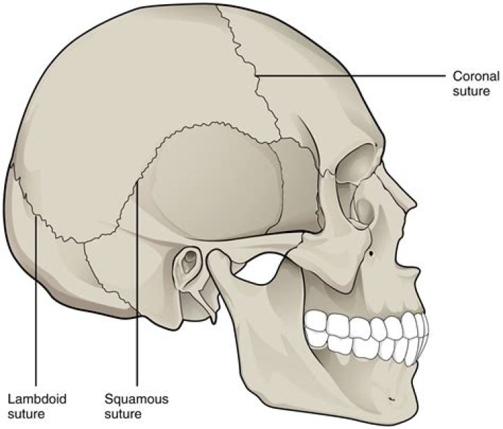
The second suture we’re going to look at is the Lambdoid suture, located at the back of the skull. It separates the occipital bone from the both the right and left parietal bones.
Where is the suture located?
A suture is a type of fibrous joint (or synarthrosis) that only occurs in the skull. The bones are bound together by Sharpey’s fibers, a matrix of connective tissue which provide a firm joint.
What is the location where the Lambdoid and sagittal sutures meet?
Posterior view The sagittal suture joins the two parietals. The lambdoid suture marks the borders between the parietal and occipital bones. The sagittal and lambdoid sutures converge into a lambda.
What does lambdoid suture mean in anatomy?
Medical Definition of lambdoid : having the Λ or λ shape of the Greek letter lambda especially : of, relating to, or being the lambda-shaped suture that connects the occipital and parietal bones.What bones form the lambdoid suture?
The lambdoid suture is a line of dense, fibrous tissue that connects the occipital bone with the parietal bones. It is continuous with the occipitomastoid suture, which connects the occipital bone with the temporal bones.
Where is the coronal suture located?
The coronal suture is a dense and fibrous association of connection tissue located in between the frontal and parietal bones of the skull. At birth, the sutures decrease in size (molding) and allow the skull to become smaller.
How do you remember the Lambdoid suture?
The word lambdoid is Greek in origin, it means “similar to lambda” – lambda is a greek letter, and it pretty much looks like an upside down V. Take a look at the suture and you can see that upside-down V-like appearance.
Why is it called Lambdoid?
Anatomical Parts The lambdoid suture (or lambdoidal suture) is a dense, fibrous connective tissue joint on the posterior aspect of the skull that connects the parietal bones with the occipital bone. It is continuous with the occipitomastoid suture. Its name comes from its lambda-like shape.Why it is called lambdoid suture?
The lambdoid suture is named due to its uppercase lambda-like shape.
How common is Lambdoid craniosynostosis?Lambdoid craniosynostosis is very rare and the only type that would cause flattening in the back of the head similar to positional plagiocephaly.
Article first time published onDoes everyone have Sutural bones?
They are found in both sexes as well as in both sides of the skull. Approximately half of Sutural bones are located in the lambdoid suture and fontanel and the masto-occipital suture. The second most common site of incidence (about 25%) is in the coronal suture. The rest occur in any remaining sutures and fontanels.
Where is the posterior fontanel located?
The one in the rear portion of the head is called the posterior fontanelle. It is triangular in shape and closes within a couple of months after birth.
Why do sutures in the skull exist?
Sutures allow the bones to move during the birth process. … This allows the bone to enlarge evenly as the brain grows and the skull expands. The result is a symmetrically shaped head.
Where is sphenoid?
The sphenoid is an unpaired bone. It sits anteriorly in the cranium, and contributes to the middle cranial fossa, the lateral wall of the skull, and the floor and sides of both orbits. It has articulations with twelve other bones: Unpaired bones – Occipital, vomer, ethmoid and frontal bones.
In which of the following bones are paranasal sinuses not found?
The skull bone that does NOT contain a paranasal sinus is the b. parietal.
What are the 5 sutures of the skull?
The main sutures of the skull are the coronal, sagittal, lambdoid and squamosal sutures.
What are the skull bones?
- Frontal bone. This is the flat bone that makes up your forehead. …
- Parietal bones. This a pair of flat bones located on either side of your head, behind the frontal bone.
- Temporal bones. …
- Occipital bone. …
- Sphenoid bone. …
- Ethmoid bone.
What is the weakest part of the skull?
Clinical significance The pterion is known as the weakest part of the skull. The anterior division of the middle meningeal artery runs underneath the pterion. Consequently, a traumatic blow to the pterion may rupture the middle meningeal artery causing an epidural haematoma.
Where is the squamous suture found?
The squamous suture connects the parietal bones, which form part of the side and top of the skull, to the temporal bones, which form part of the side and the bottom of the skull.
At what age does your skull fuse?
The sutures let the skull size grow to accommodate the baby’s growing brain. When the bones of the skull are fused together either at birth or fuse too soon, the condition is called craniosynostosis. The sutures of the skull fuse around the brain at around age 2 years.
What is the fetal skull?
1. FETAL HEAD The skull is made up of the base of skull and the vault or cranium. The vault is made of occipital bone posteriorly, the two parietals at the sides ,and the temporal bones and frontal bones anteriorly. These bones at birth are thin,easily compressible and joined by membrane.
What age does the Lambdoid suture close?
The lambdoid suture remains open during childhood, typically closing by 26 years of age, and is the most common site of wormian bones.
What is the Forum Magnum?
The foramen magnum (Latin: great hole) is a large, oval-shaped opening in the occipital bone of the skull. It is one of the several oval or circular openings (foramina) in the base of the skull. … It also transmits the accessory nerve into the skull. The foramen magnum is a very important feature in bipedal mammals.
What does the Lambdoid suture separate the parietal bones from?
2. Sagittal suture: the suture between the two parietal bones. 3. Lambdoid suture: the suture between the two parietal bones and the occipital bone.
What does the squamous suture separate?
Squamosal sutures, roughly semicircular in configuration and separate the parietal bones from the superior portion of the temporal bones. These sutures extend from the sphenoid bone anteriorly to the supra-mastoid crest posteriorly.
Is the sphenoid bone paired?
It is divided into the following parts: a median portion, known as the body of sphenoid bone, containing the sella turcica, which houses the pituitary gland as well as the paired paranasal sinuses, the sphenoidal sinuses. two greater wings on the lateral side of the body and two lesser wings from the anterior side.
What hole in the occipital bone does the spinal cord travel?
Encyclopædia Britannica, Inc. In humans the base of the cranium is the occipital bone, which has a central opening (foramen magnum) to admit the spinal cord.
How is Lambdoid craniosynostosis diagnosed?
Diagnosis. Lambdoid craniosynostosis is often diagnosed by physical exam. Your doctor may also order imaging studies to confirm the diagnosis. This is especially important with this type of craniosynostosis because it can appear similar to positional plagiocephaly, a benign condition.
Can craniosynostosis cause brain damage?
Sometimes, if the condition is not treated, the build-up of pressure in the baby’s skull can lead to problems, such as blindness, seizures, or brain damage.
Can Scaphocephaly correct itself?
They can check if it could be craniosynostosis or a common problem in babies called flat head syndrome. This is not serious and usually gets better by itself.
What is Diaphysis bone?
24013. Anatomical terminology. The diaphysis is the main or midsection (shaft) of a long bone. It is made up of cortical bone and usually contains bone marrow and adipose tissue (fat). It is a middle tubular part composed of compact bone which surrounds a central marrow cavity which contains red or yellow marrow.