Where is Crista terminalis located
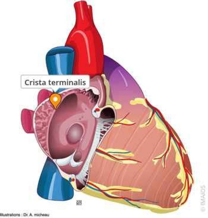
The crista terminalis (or terminal ridge) is a ridge of myocardium within the right atrium that extends along the posterolateral wall of the right atrium between the orifice of the superior vena cava to the orifice of the inferior vena cava (IVC).
Is crista terminalis in right atrium?
The crista terminalis is a variation of normal anatomical structure within the right atrium which may be misdiagnosed with an abnormal atrial mass normally visualized in the standard views on the transthoracic echocardiogram.
What is the function of the crista terminalis in the heart?
The crista terminalis acts as another anatomic conduction barrier, similar to the line of conduction block between the two venae cavae required in the animal model.
What does crista terminalis mean?
The crista terminalis is a smooth muscular ridge in the superior aspect of the right atrium, formed following resorption of the right valve of the sinus venosus.Is sulcus Terminalis the same as crista terminalis?
The terminal sulcus marks the separation of the right atrial pectinate muscles from the sinus venarum. … On the internal aspect of the right atrium, corresponding to the terminal sulcus is the crista terminalis. The superior border of the terminal sulcus designates the transverse plane in which the SA node resides.
Where is the Eustachian valve located?
The Eustachian valve (EV) is located in the superior portion of the inferior vena cava (IVC) and protrudes into the right atrial cavity. It is considered to be a functional valve in the fetus that helps direct oxygenated blood from the IVC toward the foramen ovale, thereby bypassing the pulmonary circulation.
What is Crista anatomy?
Anatomical terminology The crista ampullaris is the sensory organ of rotation. They are found in the ampullae of each of the semicircular canals of the inner ear, meaning that there are 3 pairs in total. The function of the crista ampullaris is to sense angular acceleration and deceleration.
What is an appendage of the heart?
The left atrial appendage (LAA) is a small, ear-shaped sac in the muscle wall of the left atrium (top left chamber of the heart).What does the word Crista mean?
Definition of crista : any of the inwardly projecting folds of the inner membrane of a mitochondrion.
What are the pectinate muscles and the crista terminalis?Structure. Behind the crest (crista terminalis) of the right atrium the internal surface is smooth. Pectinate muscles make up the part of the wall in front of this, the right atrial appendage. In the left atrium, the pectinate muscles are confined to the inner surface of its atrial appendage.
Article first time published onWhere does coronary sinus open?
The function of the coronary sinus is to drain the venous blood from the majority of the heart. It opens into the right atrium between the opening of inferior vena cava, the fossa ovalis and the right atrioventricular orifice.
What 3 vessels fill the right atrium?
The blood vessels include the superior and inferior vena cava. These bring blood from the body to the right atrium. Next is the pulmonary artery that carries blood from the right ventricle to the lungs.
What is tendon of Todaro?
A fibrous structure formed by the junction of the eustachian valve and the thebesian valve (valves of the inferior caval vein and coronary sinus, respectively).
Where is the sulcus Limitans?
The sulcus limitans is found in the fourth ventricle of the brain. It separates the cranial nerve motor nuclei (medial) from the sensory nuclei (lateral).
What is sulcus terminalis in tongue?
Medical Definition of sulcus terminalis 1 : a V-shaped groove separating the anterior two thirds of the tongue from the posterior third and containing the circumvallate papillae. 2 : a shallow groove on the outside of the right atrium of the heart.
What is Koch's triangle?
Koch’s triangle, named after the German pathologist and cardiologist Walter Karl Koch, is an anatomical area located in the superficial paraseptal endocardium of the right atrium, which its boundaries are the coronary sinus orifice, tendon of Todaro, and septal leaflet of the right atrioventricular valve.
Where is the Maculae located in the ear?
The vestibule is a region of the inner ear which contains the saccule and the utricle, each of which contain a macula to detect linear acceleration. Its function is to detect vertical linear acceleration. The macula of saccule lies in a nearly vertical position. It is a 2mm by 3mm patch of hair cells.
Where is the utricle and saccule located?
The utricle is a small membranous sac (part of the membranous labyrinth) and paired with the saccule lies within the vestibule of the inner ear. It has an important role in orientation and static balance, particularly in horizontal tilt.
What surrounds the Crista Galli?
The crista galli is the upper part of the perpendicular plate of the ethmoid bone of the skull. It rises above the cribriform plate. The falx cerebri (a fold of the dura mater surrounding the brain) attaches to the crista galli.
What is the function of eustachian valve?
The eustachian valve directs oxygen-rich blood from the inferior vena cava toward the foramen ovale and away from the tricuspid valve during fetal development. Ordinarily, it does not prevent reflux of right atrial blood back into the inferior vena cava because it does not function as a true valve.
Is eustachian valve present in adults?
The eustachian valve is an embryological remnant of the inferior vena cava (IVC) valve. It is usually absent or inconspicuous and has no known function in the normal adult.
Where is the Chiari network located?
The Chiari network is mobile, net-like structures occasionally seen in right atrium near the opening of inferior vena cava and coronary sinus. This is usually of no clinical significance and is often diagnosed incidentally.
What does the Cristae do in the mitochondria?
Mitochondrial cristae are the folds within the inner mitochondrial membrane. These folds allow for increased surface area in which chemical reactions, such as the redox reactions, can take place.
What is mean by Galli?
alley countable noun. An alley or alleyway is a narrow passage or street with buildings or walls on both sides. lane countable noun. A lane is a type of road, especially in the country. /gali, galI, galee, galī, gli, glI, glee, glī/
What does FOV mean in anatomy?
The field of view (FOV) is defined as the dimensions of the exact anatomic region included in a scan. In MR, the FOV may be square or asymmetric.
What is the function of the appendage?
1 Introduction. Animal appendages are external projections from the body wall that are used for very diverse functions including locomotion, grooming, and feeding.
Does everyone have a left atrial appendage?
The left atrial appendage (LAA) is a small pouch extending off the side of your left atrium in the heart that can act as a decompression chamber when atrial pressure is high. While everyone has an LAA, the size and anatomy varies, as do the issues it can cause.
What is an appendage in medical terms?
(uh-PEN-dij) In medicine, a body part (such as an arm or leg) that is attached to the main part of the body.
Is there a crista terminalis in the left atrium?
Crista terminalisInterior of the heart, frontal view (crista terminalis labeled on the left, second from the top)DetailsIdentifiersLatinCrista terminalis atrii dextri
Where are the Trabeculae Carneae located?
The trabeculae carneae (columnae carneae, or meaty ridges), are rounded or irregular muscular columns which project from the inner surface of the right and left ventricle of the heart. These are different from the pectinate muscles, which are present in the atria of the heart.
Where is the fossa Ovalis located?
The fossa ovalis is a depressed structure, of varying shapes, located in the inferior aspect of the right interatrial septum.