Where does endochondral ossification occur
Endochondral ossification occurs at two distinct sites in the vertebrate long bone – the primary (diaphyseal) and the secondary (epiphyseal) sites of ossification. Bone development initiates at the primary site.
Where does endochondral ossification typically occur?
Endochondral ossification is the process by which growing cartilage is systematically replaced by bone to form the growing skeleton. This process occurs at three main sites: the physis, the epiphysis, and the cuboidal bones of the carpus and tarsus.
Where does bone formation occur during endochondral ossification quizlet?
Terms in this set (15) Step 2 bone grows from the ossification center in linear extensions called spicules. Blood vessels grow and branch around the spicules to support the bone tissue. Endochondral ossification occurs in a cartilage model of the bone appears first in the developing embryo.
Does endochondral ossification occur in flat bones?
Endochondral ossification is the process of bone development from hyaline cartilage. All of the bones of the body, except for the flat bones of the skull, mandible, and clavicles, are formed through endochondral ossification.Where is the ossification located?
A primary ossification center is the first area of a bone to start ossifying. It usually appears during prenatal development in the central part of each developing bone. In long bones the primary centers occur in the diaphysis/shaft and in irregular bones the primary centers occur usually in the body of the bone.
How does endochondral ossification form bones?
Endochondral Ossification This process involves the replacement of hyaline cartilage with bone. It begins when mesoderm-derived mesenchymal cells differentiate into chondrocytes. Chondrocytes proliferate rapidly and secrete an extracellular matrix to form the cartilage model for bone.
What is the primary ossification center in endochondral ossification?
Primary center of ossification The perichondrium becomes the periosteum. The periosteum contains a layer of undifferentiated cells (osteoprogenitor cells) which later become osteoblasts. The osteoblasts secrete osteoid against the shaft of the cartilage model (Appositional Growth).
Where is the primary ossification center during endochondral ossification in a long bone?
The first site of ossification occurs in the primary center of ossification, which is in the middle of diaphysis (shaft). The perichondrium becomes the periosteum.Where does bone formation occur during endochondral ossification multiple choice question?
Bones at the base of the skull and long bones form via endochondral ossification. In a long bone, for example, at about 6 to 8 weeks after conception, some of the mesenchymal cells differentiate into chondroblasts (cartilage cells) that form the hyaline cartilaginous skeletal precursor of the bones (Figure 6.4.
Are large phagocytic cells found in bone?Osteoclasts. Osteoclasts are large multinucleated phagocytic cells derived from the macrophage-monocyte cell lineage (23). They migrate from bone marrow to a specific skeletal site.
Article first time published onWhat is endochondral ossification quizlet?
Endochondral ossification. –a process whereby cartilage is replaced by bone. -forms both compact and spongy bone. Only $35.99/year. Method used in the formation of most bones, especially long bones.
Does endochondral ossification occurs within fibrous connective tissue membranes?
Endochondral ossification occurs within fibrous connective tissue membranes. Endochondral ossification leads to the formation of the clavicles and cranial bones. … It is more complex than intramembranous ossification because the hyaline cartilage must be broken down as ossification proceeds.
Where does bone formation occur during endochondral ossification hyaline cartilage model fibrous membranous sheet adipose tissue?
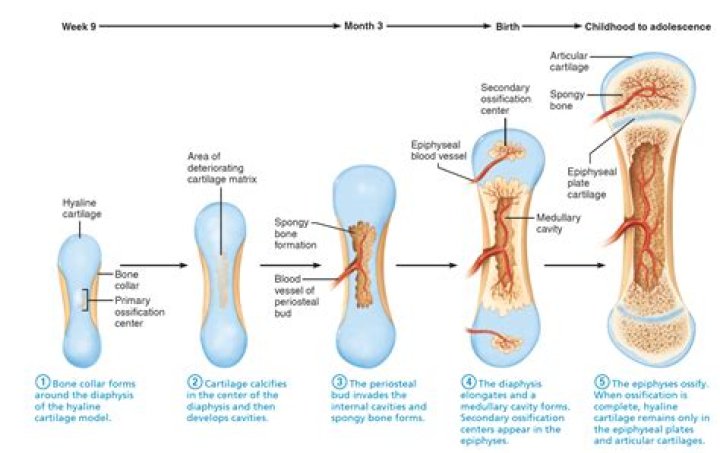
First, the bone collar forms around the diaphysis of the hyaline cartilage model. Then, cartilage in the center of the diaphysis calcifies and develops cavities.
In what ways do Intramembranous and endochondral ossification differ?
The main difference between endochondral ossification and intramembranous ossification is that the endochondral ossification is the method of forming a bone through a cartilage intermediate while the intramembranous ossification directly forms the bone on the mesenchyme.
What are the steps in the process of endochondral ossification?
- Cartilage enlarges; Chondrocytes die.
- blood vessels grow into perichondrium; cells convert to osteoblasts; shaft becomes covered with superficial bone.
- more blood supply and osteoblasts; produces spongy bone; formation spreads on shaft.
- Osteoclasts create medullary cavity; appositional growth.
What is step 4 in the endochondral ossification process?
4) Blood vessels grow around the edges of the cartilage. 5) Perichondrial cells become osteoblasts and produce a superficial layer of bone.
In which zone of endochondral ossification do chondrocytes undergo mitosis?
This zone contains normal, resting hyaline cartilage. Zone of proliferation. In this zone, chondrocytes undergo rapid mitosis, forming distinctive looking stacks.
How does endochondral ossification occur?
Endochondral ossification involves the replacement of hyaline cartilage with bony tissue. … At the same time, the cartilage in the center of the diaphysis begins to disintegrate. Osteoblasts penetrate the disintegrating cartilage and replace it with spongy bone. This forms a primary ossification center.
Where does Endochondral lengthening occur?
Lengthening of the bone occurs in the epiphyseal growth plate. Cells on the metaphyseal side ossify (zone of provisional calcification), and cells on the epiphyseal side multiply (zone of proliferation).
Where does bone formation occur in endochondral ossification?
Bones at the base of the skull and long bones form via endochondral ossification. In a long bone, for example, at about 6 to 8 weeks after conception, some of the mesenchymal cells differentiate into chondrocytes (cartilage cells) that form the cartilaginous skeletal precursor of the bones ((Figure)a).
What happens to chondrocytes during endochondral ossification?
The process of endochondral ossification. (a) During endochondral ossification, mesenchymal cells differentiate into chondrocytes and lead to the formation of cartilage templates. Vascularization occurs around these templates, and osteoblasts differentiate around the central area in the bone collar.
When cartilage is produced at the epiphyseal side?
The epiphyseal plate, the area of growth composed of four zones, is where cartilage is formed on the epiphyseal side while cartilage is ossified on the diaphyseal side, thereby lengthening the bone.
Where within the epiphyseal plate are the dividing cartilage cells located?
Near the outer end of each epiphyseal plate is the active zone dividing the cartilage cells.
What are the zones in endochondral bone ossification?
In endochondral ossification: The bone is formed onto a temporary cartilage model. The cartilage model grows (zone of proliferation), then chondrocytes mature (zone of maturation) and hypertropy (zone of hypertrophy), and growing cartilage model starts to calcify.
Where are the primary and secondary ossification centers located in the long bone during endochondral ossification?
Each of these bones has a primary center of ossification. The zone of endochondral ossification spreads from the primary ossification center toward the ends of the cartilage. These slides do not show secondary ossification centers. Note the bone of the diaphysis.
Where is the primary center of ossification in endochondral ossification of a long bone quizlet?
In long bones the primary centers occur in the diaphysis/shaft and in irregular bones the primary centers occur usually in the body of the bone. The site where bone formation continues after beginning in the long shaft or body of the bone, usually in an epiphysis; secondary ossification center.
Is Endochondral an ossification?
Endochondral ossification is the process by which the embryonic cartilaginous model of most bones contributes to longitudinal growth and is gradually replaced by bone.
What is phagocytic cell?
phagocyte, type of cell that has the ability to ingest, and sometimes digest, foreign particles, such as bacteria, carbon, dust, or dye. … In the blood, two types of white blood cells, neutrophilic leukocytes (microphages) and monocytes (macrophages), are phagocytic.
Which cell is phagocytic in nature?
In humans, and in vertebrates generally, the most-effective phagocytic cells are two kinds of white blood cells: the macrophages (large phagocytic cells) and the neutrophils (a type of granulocyte).
What tissue forms the model for endochondral ossification quizlet?
Endochondral ossification converts hyaline cartilage “bone” models into true bones (i.e., hyaline cartilage serves as a template for bone formation). Endochondral ossification occurs within fibrous connective tissue membranes.
What type of ossification occurs in the skull?
The direct conversion of mesenchymal tissue into bone is called intramembranous ossification. This process occurs primarily in the bones of the skull. In other cases, the mesenchymal cells differentiate into cartilage, and this cartilage is later replaced by bone.