What is VRG and DRG
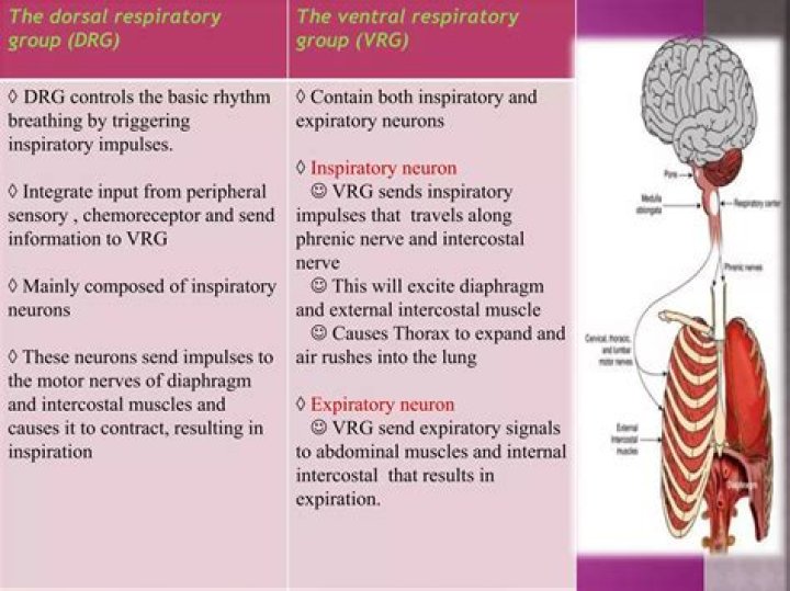
The automatic rhythm of breathing is generated by specialized neurons of the medulla oblongata: the Dorsal Respiratory Group
What is the VRG?
The VRG is a ventrolateral column of respiratory neurons that includes the nucleus ambiguous, nucleus retroambigulalis, and the pre-Bötzinger and Bötzinger complexes. … The Bötzinger complex is located at the most rostral end of the VRG and contains neurons that inhibit most inspiratory neurons during expiration.
What is DRG in respiratory?
The dorsal respiratory group (DRG) has the most fundamental role in the control of respiration, initiating inspiration (inhalation). The DRG is a collection of neurons forming an elongated mass that extends most of the length of the dorsal medulla.
Where are DRG and VRG located?
Other Respiratory Centers Other anatomical or functional groups of neurons involved in control of ventilation include the ventral and dorsal respiratory groups (VRG and DRG) in the medulla. The ventral respiratory group (VRG) is a column of neurons that fire action potentials in phase with respiration.What is Apneustic?
The apneustic center, which is located in the lower pons, is thought to excite the inspiratory center. Rather than abruptly sending signals to the inspiratory muscles to contract, stimulation of the apneustic center leads to a gradual increase in the firing rate of the inspiratory muscles.
How does Pons control breathing?
The pons is the other respiratory center and is located underneath the medulla. Its main function is to control the rate or speed of involuntary respiration. It has two main functional regions that perform this role: The apneustic center sends signals for inspiration for long and deep breaths.
How does DRG differ from VRG?
Dorsal respiratory groups (DRG) DRG nerves extend into the VRG, but the VRG neurons do not extend into the DRG. Vagus and glossopharyngeal nerves bring sensory impulses to the DRG from the lungs, airways, peripheral chemoreceptors, and joint proprioceptors. Input modifies the breathing pattern.
Where are the I and E neurons located?
I and E neurons were further classified into augmenting, decrementing, and other types based on their firing patterns. Almost all the respiratory neurons recorded were located around the nucleus ambiguus and the nucleus retroambigualis, corresponding to the ventral respiratory group (VRG) of the cat.How is breathing regulated in human?
Breathing occurs due to repeated contractions of a large muscle called the diaphragm. The rate of breathing is regulated by the brain stem. It monitors the level of carbon dioxide in the blood and triggers faster or slower breathing as needed to keep the level within a narrow range.
What is the result of inspiration?Inspiration (inhalation) is the process of taking air into the lungs. It is the active phase of ventilation because it is the result of muscle contraction. During inspiration, the diaphragm contracts and the thoracic cavity increases in volume. This decreases the intraalveolar pressure so that air flows into the lungs.
Article first time published onWhere is the medulla located?
medulla oblongata, also called medulla, the lowest part of the brain and the lowest portion of the brainstem. The medulla oblongata is connected by the pons to the midbrain and is continuous posteriorly with the spinal cord, with which it merges at the opening (foramen magnum) at the base of the skull.
What muscles would the ventral respiratory group VRG influence?
The medulla oblongata contains the dorsal respiratory group (DRG) and the ventral respiratory group (VRG). The DRG is involved in maintaining a constant breathing rhythm by stimulating the diaphragm and intercostal muscles to contract, resulting in inspiration.
What causes Kussmaul breathing?
Causes: Kussmaul breathing is usually caused by high acidity levels in the blood. Cheyne-Stokes breathing is usually related to heart failure, stroke, head injuries, or brain conditions. Pattern: Kussmaul breathing doesn’t alternate between periods of fast and slow breathing.
What directly stimulates the central Chemoreceptors?
What directly stimulates the central chemoreceptors, thus increasing respiration? H+ (hydrogen ions). CO2 is converted to H+ in the extracellular fluid of the brain.
What is the pons main function?
The pons is part of a highway-like structure between the brain and the body known as the brainstem. The brainstem is made up of three sections, and carries vital information to the body. The pons relays information about motor function, sensation, eye movement, hearing, taste, and more.
What is the role of the pons?
The pons, while involved in the regulation of functions carried out by the cranial nerves it houses, works together with the medulla oblongata to serve an especially critical role in generating the respiratory rhythm of breathing. Active functioning of the pons may also be fundamental to rapid eye movement (REM) sleep.
What does the pons regulate?
The pons contains nuclei that relay signals from the forebrain to the cerebellum, along with nuclei that deal primarily with sleep, respiration, swallowing, bladder control, hearing, equilibrium, taste, eye movement, facial expressions, facial sensation, and posture.
What regulates breathing and heartbeat?
The cerebellum sits at the back of your head, under the cerebrum. It controls coordination and balance. The brain stem sits beneath your cerebrum in front of your cerebellum. It connects the brain to the spinal cord and controls automatic functions such as breathing, digestion, heart rate and blood pressure.
What part of the brain regulates breathing?
Medulla. At the bottom of the brainstem, the medulla is where the brain meets the spinal cord. The medulla is essential to survival. Functions of the medulla regulate many bodily activities, including heart rhythm, breathing, blood flow, and oxygen and carbon dioxide levels.
What are the 2 types of motor neurons?
Motor neurons are a specialized type of brain cell called neurons located within the spinal cord and the brain. They come in two main subtypes, namely the upper motor neurons and the lower motor neurons. The upper motor neurons originate in the brain and travel downward to connect with the lower motor neurons.
What is the role of neural pools in the CNS?
The billions of neurons in the CNS are organized into neuronal pools. These functional groups of neurons integrate incoming information from receptors or different neuronal pools and then forward the processed information to other destinations.
Are neuron cells?
A neuron or nerve cell is an electrically excitable cell that communicates with other cells via specialized connections called synapses. It is the main component of nervous tissue in all animals except sponges and placozoa. … A typical neuron consists of a cell body (soma), dendrites, and a single axon.
What is the true meaning of inspiration?
1 : an inspiring agent or influence. 2a : the quality or state of being inspired. b : something that is inspired a scheme that was pure inspiration. 3 : the act of drawing in specifically : the drawing of air into the lungs.
What is the difference between inspiration and expiration?
The difference between inspiration and expiration is, the inspiration is an active process where it brings air into the lungs while expiration is a passive process, which is the expulsion of the air out of the lungs.
Which conditions are correct for inspiration?
Inspiration occurs when intrapulmonary pressure falls below atmospheric pressure, and air moves into the lungs. Intrapulmonary pressure falls below atmospheric pressure when the diaphragm contracts and increases the thoracic volume. The diaphragm is the major inspiratory muscle.
Can you live without medulla?
Making up a tail-like structure at the base of the brain, the medulla oblongata connects the brain to the spinal cord, and includes a number of specialized structures and functions. While every part of the brain important in its own way, life cannot be sustained without the work of the medulla oblongata.
What is medulla in kidney?
The renal medulla is the innermost part of the kidney. The renal medulla is split up into a number of sections, known as the renal pyramids. … The renal medulla (Latin: medulla renis ‘marrow of the kidney’) contains the structures of the nephrons responsible for maintaining the salt and water balance of the blood.
Why is the medulla important for our survival?
The medulla contains the nuclei that control vital (survival) functions: the respiratory and cardiovascular centres, swallowing, blood pressure and vomiting (Box 6.3). A key nucleus involved in these functions is the nucleus of the solitary tract (NTS).
What happens physiologically during breathing?
When you breathe in, or inhale, your diaphragm contracts and moves downward. This increases the space in your chest cavity, and your lungs expand into it. The muscles between your ribs also help enlarge the chest cavity. They contract to pull your rib cage both upward and outward when you inhale.
What causes exhalation?
The process of exhalation occurs due to an elastic recoil of the lung tissue which causes a decrease in volume, resulting in increased pressure in comparison to the atmosphere; thus, air rushes out of the airway. There is no contraction of muscles during exhalation; it is considered a passive process.
What stimulates the respiratory center?
An increased concentration of carbon dioxide normally stimulates the body’s respiratory center in the medulla, and to a lesser extent, by decreased levels of oxygen in arterial blood.