What is indirect immunofluorescence assay
Indirect immunofluorescence assay: A laboratory test used to detect antibodies in serum or other body fluid. The specific antibodies are labeled with a compound that makes them glow an apple-green color when observed microscopically under ultraviolet light.
What is the difference between direct and indirect immunofluorescence?
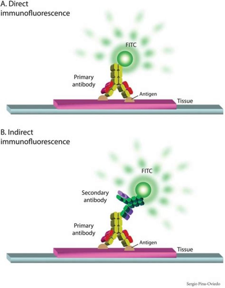
The key difference between direct and indirect immunofluorescence is that the direct immunofluorescence uses a single antibody that works against the target of interest while the indirect immunofluorescence uses two antibodies to label the target of interest.
What is the direct and the indirect method of immunofluorescence?
Immunofluorescence staining (IF) can be performed in two ways, by direct or indirect detection. Direct IF uses a dye-conjugated antibody to stain the target protein. Indirect IF involves first binding the primary antibody to the target, then detecting the primary antibody using a conjugated secondary antibody.
What is immunofluorescence used for?
Immunofluorescence is commonly used in molecular and cell biology labs as a robust and simple method to reliably localize molecules on a wide range of fixed cells or tissues.How does immunofluorescence assay work?
Immunofluorescence assay (IFA) is a standard virologic technique to identify the presence of antibodies by their specific ability to react with viral antigens expressed in infected cells; bound antibodies are visualized by incubation with fluorescently labeled antihuman antibody.
What is indirect ELISA?
Indirect ELISA is a two-step ELISA which involves two binding process of primary antibody and labeled secondary antibody. The primary antibody is incubated with the antigen followed by the incubation with the secondary antibody. … Samples with antibodies are added and washed.
What is direct and indirect assay?
Direct ELISAs use a conjugated primary antibody, while indirect ELISAs include an additional amplification step. In an indirect ELISA, an unconjugated primary antibody binds to the antigen, then a labeled secondary antibody directed against the host species of the primary antibody binds to the primary antibody.
Which microscope is used in immunofluorescence analysis?
Several microscope designs can be used for analysis of immunofluorescence samples; the simplest is the epifluorescence microscope, and the confocal microscope is also widely used. Various super-resolution microscope designs that are capable of much higher resolution can also be used.What is immunofluorescence and its types?
Immunofluorescence (IF) is a type of immunohistochemistry technique that utilizes fluorophores to visualize various cellular antigens such as proteins.
What is immunoblot assay?Immunoblotting (western blotting) is a rapid and sensitive assay for the detection and characterization of proteins that works by exploiting the specificity inherent in antigen-antibody recognition.
Article first time published onIs indirect immunofluorescence quantitative?
These biological standards can be routinely used as internal references to establish the quantitative phenotype of lymphoid cells. The present method is referred to as quantitative indirect immunofluorescence assay (QIIF). It can be used with any flow cytometer equipped with a microcomputer.
What is the difference between direct and indirect immunohistochemistry?
In direct detection methods, the primary antibody is directly conjugated to a label. During indirect detection, the primary antibody is bound by a labeled secondary antibody that has been raised against the host species of the primary antibody.
What is direct Elisa?
A direct ELISA (enzyme-linked immunosorbent assay) is a plate-based immunosorbent assay intended for the detection and quantification of a specific analyte (e.g. antigens, antibodies, proteins, hormones, peptides, etc.) from within a complex biological sample.
What is indirect fluorescent antibody test?
The indirect fluorescent antibody test (IFA) is a semi-quantitative, sensitive, and rapid test for the detection of anti-rabies virus (RABV) immunoglobulin M (IgM) and G (IgG) antibodies in serum and cerebral spinal fluid (CSF) samples.
What is IFA in microbiology?
Indirect fluorescent antibody (IFA) tests (Figure 2) are used to look for antibodies in patient serum. For example, an IFA test for the diagnosis of syphilis uses T. pallidum cells isolated from a lab animal (the bacteria cannot be grown on lab media) and a smear prepared on a glass slide.
What is indirect assay?
Indirect ELISA Assay Indirect ELISA is a two-step binding process involving the use of a primary antibody and a labeled secondary antibody. In this method, the primary antibody is incubated with the antigen-coated wells. Next, a labeled secondary antibody that recognizes the primary antibody is added.
Why is it called indirect ELISA?
The detection antibody can be enzyme conjugated, in which case this is referred to as a direct sandwich ELISA. If the detection antibody used is unlabeled, a secondary enzyme-conjugated detection antibody is required. This is known as an indirect sandwich ELISA.
When is indirect ELISA used?
The indirect ELISA is used for the quantitative estimation of antibodies in the serum and other body fluids. In this method, specimens are added to microtiter plate wells coated with antigen to which specific antibodies are to be detected. After a period of incubation, the wells are washed.
What are the differences between direct and indirect ELISA?
The major difference between direct and indirect ELISA is that only one antibody is used in direct ELISA, while indirect ELISA requires two antibodies. … Meanwhile, different enzyme-substrate detection systems can be used for the same primary antibody, allowing for fine-tuning and improving the ELISA protocol.
What is the difference between indirect and competitive ELISA?
Competitive ELISA The steps of a competitive ELISA are different from those used in indirect and sandwich ELISA, with the main difference being the competitive binding step between the sample antigen and the “add-in” antigen. The sample antigen is incubated with the unlabeled primary antibody.
Which type of ELISA is best?
AdvantagesSandwich ELISAHigh flexibility. High sensitivity. High specificity, since different antibodies bind to the same antigen for detection.Competitive ELISAHigh flexibility. High sensitivity. Best for the detection of small antigens, even when they are present in low concentrations.
Can you combine direct and indirect immunofluorescence?
If for double staining using monoclonal antibodies of the same species only one antibody is conjugated with FITC or TRITC, a combination of indirect and direct immunofluorescence is possible.
What is indirect Labelling?
Direct labelling (left) uses an antibody tagged with a fluorescent marker to label target antigens while indirect labelling (right) uses secondary antibodies tagged with the markers to target the primary antibody that has bound to the target antigen.
What are immunofluorescence techniques?
The immunofluorescence is a histochemical laboratory staining technique that uses the specificity of Abs to their antigen. It is a widely used in immunohistochemistry based on the use of some fluorochromes [5] to visualize the location of the Abs.
What is the difference between immunofluorescence and immunohistochemistry?
immunofluorescence is commonly used to stain microbiological cells. immunohistochemistry is commonly used to stain sections of biological tissue.
Is immunofluorescence a type of immunohistochemistry?
Let us put it another way,immunohistochemistry and immunocytochemistry are one type of immunofluorescence. … Immunocytochemistry is performed on sample of intact cells. Immunofluorescence may be used to analyze the distribution of proteins, glycans, and small biological and non-biological molecules in cells or tissues.
What can immunofluorescent staining tell you?
Immunofluorescence Staining Immunofluorescence (IF) staining uses tissue sections or cultured cell lines as an antigenic source and detects the specific recognition of autoantibodies to native autoantigens on fixed cells/tissues (Figure 6.1A).
What is the difference between immunoblot and Western blot?
In a Western Blot, proteins are separated by size. Therefore, more than one protein can be present at any one position (e.g. at 31kDa, Osp A and non-specific antigen are present.) Whereas on an ImmunoBlot, pure proteins are sprayed at specific positions on the blot.
What is the difference between immunoblotting and immunoprecipitation?
Immunoprecipitation has long been used as a tool for assessing a protein’s presence, while immunoblotting provides a more accurate means of quantitating the amount of the protein that is present. Immunoblotting also serves to establish or verify the identity of proteins isolated in immunoprecipitation.
When is immunoblotting used?
The western blot (sometimes called the protein immunoblot), or western blotting, is a widely used analytical technique in molecular biology and immunogenetics to detect specific proteins in a sample of tissue homogenate or extract.
What is an IFAT test?
The immunofluorescence antibody test (IFAT) is one of the most commonly used techniques for detection of anti-Leishmania antibodies. The purpose of this study was to assess whether there is a correlation between clinical signs and IFAT titers in dogs naturally infected with Leishmania.