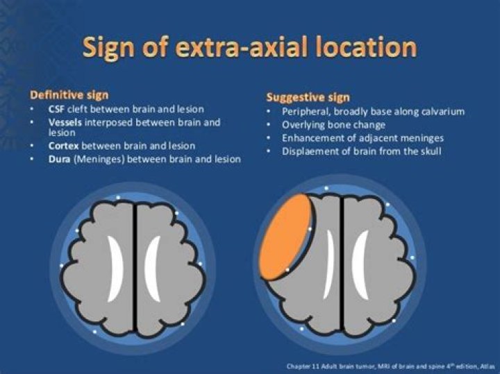
What is an extra axial lesion
Extra-axial tumors are lesions, neoplastic and not, which are external to the brain parenchyma and can originate in the skull, meninges, cranial nerves, and brain appendages such as the pituitary gland. Surgery provides a diagnosis and can be the first step in the treatment.
What is extra-axial meningioma?
Extra-axial brain tumors are the most common adult intracranial neoplasms and encompass a broad spectrum of pathologic subtypes. Meningiomas are the most common extra-axial brain tumor (approximately one-third of all intracranial neoplasms) and typically present as slowly growing dural-based masses.
What is intra and extra-axial?
The intra-axial tumors are located within brain parenchyma and arise from the brain cells, while the extra-axial tumors are located outside brain parenchyma and arise from structures lining the brain or surrounding it.
What is extra-axial space in the brain?
The brain is surrounded by cerebrospinal fluid (CSF) within the sulci, fissures and basal cisterns. CSF is also found centrally within the ventricles. The sulci, fissures, basal cisterns and ventricles together form the ‘CSF spaces‘, also known as the ‘extra-axial spaces’.What is an extra-axial calcification?
Extra-axial brain stones comprise tumors and exaggerated physiological calcifications. Intra-axial calcifications can be classified as vascular, neoplastic, congenital, infectious, and endocrine/metabolic etiologies. Vascular causes represent a substantial portion of intra-axial lesions.
At what size should a meningioma be removed?
Ideally, surgical removal of meningioma entails removal of a one-centimeter margin all the way around the tumor. However, this type of resection is not always possible, especially in the skull base. These deep-seated tumors in the skull base require referral to a skull base neurosurgeon.
What is the difference between a brain tumor and a brain lesion?
A brain tumor is a specific type of brain lesion. A lesion describes any area of damaged tissue. All tumors are lesions, but not all lesions are tumors. Other brain lesions can be caused by stroke, injury, encephalitis and arteriovenous malformation.
What is a lesion on the brain?
A brain lesion is an abnormality seen on a brain-imaging test, such as magnetic resonance imaging (MRI) or computerized tomography (CT). On CT or MRI scans, brain lesions appear as dark or light spots that don’t look like normal brain tissue.Can you see a CSF leak on a CT scan?
CT myelography. This test is considered the gold standard for diagnosing and locating CSF leaks. It uses a CT scan and a contrast dye to locate CSF leaks anywhere in the skull base. It provides the most precise location of a CSF leak and helps to determine the most appropriate treatment plan.
What causes extra-axial fluid in adults?Hydrocephalus ex-vacuo occurs when a stroke or injury damages the brain and brain matter actually shrinks. The brain may shrink in older patients or those with Alzheimer’s disease, and CSF volume increases to fill the extra space. In these instances, the ventricles are enlarged, but the pressure usually is normal.
Article first time published onCan meningioma be intra axial?
Most intraparenchymal meningiomas are of fibrous type, whereas the extra-axial meningiomas are of the meningothelial type. The most common location is the frontal lobe followed by the parietal lobe. The imaging findings of intraparenchymal meningioma are nonspecific and most cases are misdiagnosed preoperatively. ].
What is intra axial tumor?
Brain tumors that are rooted in the brain Parenchyma are referred to as intra-axial. If the origin of the tumor is outside of the brain (or it is due to metastasis), it is called extra-axial (4, 5).
Can brain calcification cause headaches?
Clinically, brain calcification can include symptons such as migraine, parkinsonism, psychosis or dementia.
Can calcium deposits on your brain be serious?
While considered by many to be benign, these calcium phosphate deposits or “brain stones” can become large and are associated with neurological symptoms that range from seizures to parkinsonian symptoms.
What are the symptoms of calcification of the brain?
Muscle cramping (dystonia), uncontrollable spasmodic irregular movements (chorea), and seizures can also occur. Occasional symptoms include sensory changes, headaches and urinary incontinence. Associated symptoms include loss of contact with reality (psychosis), mood swings and loss of acquired motor skills.
Can you tell if a brain tumor is cancerous from an MRI?
There is no way to tell from symptoms alone if a tumor is benign or malignant. Often an MRI scan can reveal the tumor type, but in many cases, a biopsy is required.
Are brain lesions serious?
So a brain lesion is an area of injury or disease within the brain. While the definition sounds simple, understanding brain lesions can be complicated. That’s because there are many types of brain lesions. They can range from small to large, from few to many, from relatively harmless to life threatening.
Can a brain lesion be nothing?
Lesions can be due to disease, trauma or a birth defect. Sometimes lesions appear in a specific area of the brain. At other times, the lesions are present in a large part of the brain tissue. At first, brain lesions may not produce any symptoms.
How serious is meningioma surgery?
Complications of surgery — Possible complications of surgery include damage to nearby normal brain tissue, bleeding, spinal fluid leakage, and infection. Potentially serious complications can include: Temporary accumulation of fluid in the brain (cerebral edema) is common after surgery for meningiomas.
How long do you stay in the hospital after meningioma surgery?
The hospital stay after surgery for a meningioma can range from a few days to a couple of weeks, depending on how large the tumor is, where it’s located, and the type of procedure used to remove it.
What happens if you don't remove a meningioma?
In about 95 percent of recurrences, the new meningioma grows in the same spot as before. In some cases, total resection, or removal, is not possible. If a meningioma tumor is not removed completely, it is likely to regrow within 10 to 20 years.
What kind of doctor treats CSF leak?
Our sinus specialists, otologists/neurotologists, and neurosurgeons collaborate to diagnose and treat cranial CSF leaks. These are most often treated with surgery.
Does CSF leak make you tired?
Any CSF leak is most often characterized by orthostatic headaches, which worsen when standing, and improve when lying down. Other symptoms can include neck pain or stiffness, nausea, vomiting, dizziness, fatigue, and a metallic taste in the mouth.
How do they fix a CSF leak?
Surgery is often the best treatment option for cranial CSF leaks. Surgical approaches are tailored to the exact location of the leak. Once surgeons reach the leak site, they repair the hole by plugging it with tissue or fat.
Can Brain Lesions be caused by stress?
Psychological stress is linked to multiple sclerosis (MS) severity (e.g., to a heightened risk of brain lesion development).
Do brain lesions cause headaches?
Brain lesions don’t appear to cause any long-term damage. Two large studies found people with migraines didn’t have any more changes to their brain function or thinking than those who don’t get the headaches. One study concluded these lesions don’t affect your brain health.
Is fluid on the brain serious?
Hydrocephalus is a build-up of fluid in the brain. The excess fluid puts pressure on the brain, which can damage it. If left untreated, hydrocephalus can be fatal.
Can you live a normal life with a brain shunt?
Overview. Many people with normal pressure hydrocephalus enjoy a normal life with the help of a shunt. Regular, ongoing checkups with the neurosurgeon will help ensure that your shunt is working correctly, your progress is on track, and you are free to keep living the way you want.
What will happen if hydrocephalus is not treated?
Without treatment, hydrocephalus results in compromised mental functioning, visual disturbances, walking difficulty, incontinence, and reduced conscious state.
What does a meningioma look like on MRI?
Meningiomas typically appear as lobular, extra axial masses with well-circumscribed margins (Fig. 5a). They typically have a broad-based dural attachment and, if sufficiently large, inward displacement of the cortical grey matter [5].
How do they remove a meningioma?
The most common type of surgery to remove a meningioma is called a craniotomy. This procedure involves making an incision in the scalp and removing a piece of bone from the skull. The neurosurgeon can then access and remove the tumor, or as much of the tumor as possible without risk of severe damage to the brain.