What is a CT temporal bones
Temporal bone CT is a limited kind of head CT that focuses on the lower part of the skull and the surrounding soft tissues, and is often used in patients with hearing loss, chronic ear infections, and middle and inner ear diseases.
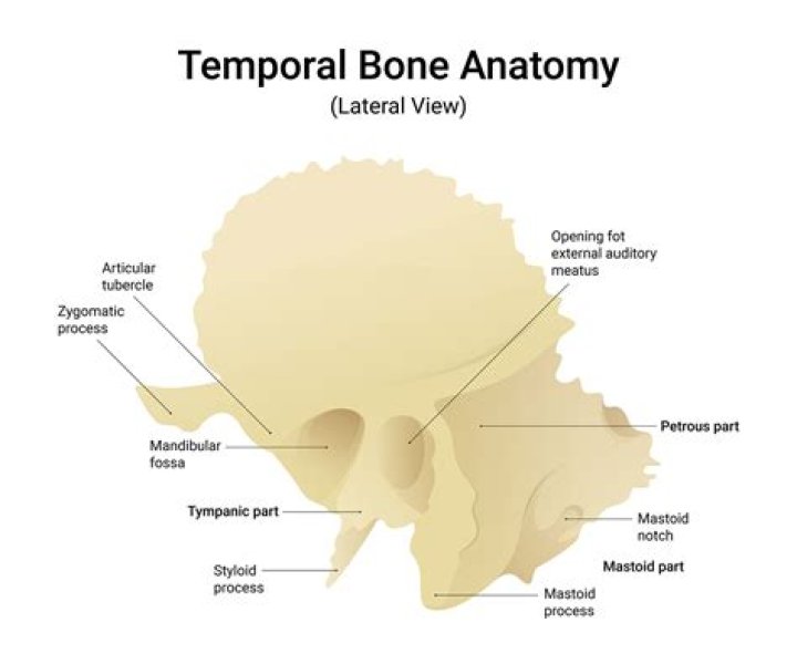
What are temporal bones?
The temporal bones are paired bones that help make up the sides and base of the skull (cranium). This places them lateral—to the side of—the temporal lobes of the brain’s cerebral cortex, ensuring that the cranium is properly supported and protecting the important structures there.
What is HRCT temporal bone CT scan?
HRCT temporal bone axial scan is a high resolution computed tomography (HRCT) imaging test which helps to visualise specially the soft tissues and bones of the temporal region of the brain (middle ear).
Is a CT scan of temporal bone safe?
Note that most temporal bone CT scans are “high radiation” procedures because enough Xray energy must be used to “see” into a very hard bone (temporal bone). All Xrays increase cancer risk. Accordingly, CT scans should not be done to “screen” for SCD. Safe tests such as the VEMP should be used first.What does CT facial bones show?
A CT of your facial bones can help your physician to assess the possible causes of such things as headaches, seizures, dizziness or swelling. It can also be used to examine other possible problems, such as those from an injury or from a tumor.
What muscles are attached to the temporal bone?
Muscular attachments The temporalis muscle originates from the temporal fossa, which is formed partially by the lateral aspect of the temporal bone. The sternocleidomastoid, splenius capitis, longissimus capitis and digastric are all attached to the mastoid process of the temporal bone.
Why is it called temporal bone?
Etymology. Its exact etymology is unknown. It is thought to be from the Old French temporal meaning “earthly,” which is directly from the Latin tempus meaning “time, proper time or season.” Temporal bones are situated on the sides of the skull, where grey hairs usually appear early on.
What causes the temporal bone to hurt?
Temporal bone trauma is usually the result of blunt head injury and patients commonly suffer from multiple other body injuries. Motor vehicle accidents are the most common cause, with falls and gunshot wounds contributing to a lesser extent.How long does a temporal bone CT take?
Depending on the reason for your test, the procedure can take anywhere from 10 to 30 minutes. You will receive the results of the exam from your doctor.
Can a CT scan detect inner ear problems?CT scans use electromagnetic radiation to take a series of X-rays of the interior structures of the ear and create a computerized three-dimensional image. CT scans may reveal damage to the bony components of the ear or an abnormal bone growth in the middle ear, a condition called otosclerosis.
Article first time published onWhy use HRCT temporal bone?
HRCT temporal bone is an efficacious modality for accurate delineation of the anatomy and pathological involvement of temporal bone. We can hence conclude HRCT is useful for diagnosis, surgical planning and management of temporal bone pathologies.
Which of the following is the clinical indication of HRCT temporal bone?
These clinical conditions are hearing loss, external auditory canal atresia, middle ear inflammation/cholesteatoma, temporal bone trauma, pulsatile tinnitus and vascular tympanic membrane.
Does CT head show sinuses?
Most CT scans of the head do not . include all of the sinuses. For most problems in the brain that cause headache, MRI scans are more sensitive. For the detection of a recent brain hemorrhage or for sinus disease, CT is more helpful.
Can you see inflammation on a head CT scan?
CT scans can show if there is swelling or bleeding in the brain or a fracture in the skull. If you have signs of a serious injury, a CT scan is usually the best first test to diagnose it.
Does CT head include orbits?
CT scanning of the sinuses, orbits, or face uses a thin beam of X-ray and a rapidly moving X-ray tube to acquire data from different angles around your head, which is used to create cross sectional images.
What does a temporal bone fracture feel like?
Temporal bone fractures, especially the oblique variety (see above), may impair hearing and cause dizziness. There often is blood seen behind the ear-drum (hemotympanum). Either a conductive or sensorineural hearing loss or both may be present.
How serious is a temporal bone fracture?
As stated right from the first paragraph, temporal bone fractures cause several serious complications. These include facial nerve injury, CSF leak, SNHL, conductive hearing loss (CHL), cholesteatoma formation, and stenosis of the ear canal.
Is temporal bone a facial bone?
The bones that make up the neurocranium are the singular occipital, frontal, sphenoid, and ethmoid bones, and the paired temporal and parietal bones. The neurocranium is, therefore, composed of eight bones. As we have already seen, the facial bones sometimes include the sphenoid and ethmoid bones, and sometimes not.
Which bone is anterior to the temporal bone?
The squamous part of the temporal bone also articulates with the sphenoid bone anteriorly and the parietal bone laterally. The zygomatic process of the temporal bone also articulates with the zygomatic bone to form the zygomatic arch (i.e. cheekbones).
What bone is superior to the temporal bone?
The temporal bone articulates anteriorly with the sphenoid bone, above with the parietal bone, and posteriorly with the occipital bone.
Does the temporal bone have a sinus?
Explanation: There are four paranasal sinuses in the head: the frontal, maxillary, sphenoid, and ethmoid sinuses. They function in lightening the skull, and creating mucous for the nasal cavity. The temporal bone does not contain a sinus.
What's better CT or MRI?
Both MRIs and CT scans can view internal body structures. However, a CT scan is faster and can provide pictures of tissues, organs, and skeletal structure. An MRI is highly adept at capturing images that help doctors determine if there are abnormal tissues within the body. MRIs are more detailed in their images.
What is the procedure for CT scan?
During a CT scan, you lie in a tunnel-like machine while the inside of the machine rotates and takes a series of X-rays from different angles. These pictures are then sent to a computer, where they’re combined to create images of slices, or cross-sections, of the body.
How much does a CT scan of the ear cost?
ProcedurePrice RangeCT Bone Density Scan Cost Average$300 – $3,800CT Ear Cost Average$350 – $7,700CT Maxillofacial (Sinus) Cost Average$600 – $6,000CT Neck Cost Average$1,000 – $7,600
Can temporal bone cause deafness?
A temporal bone fracture may cause facial paralysis, hearing loss, bruising behind the ear, and bleeding from the ear.
How long does a temporal fracture take to heal?
It could take a month or more to fully heal. It may take 6-8 weeks for the bruising around the temporal nerve to go away. A repeat hearing test and follow-up with Ear, Nose and Throat (ENT) clinic may be required after you have healed.
How do you manage Hemotympanum?
The treatment of an uncomplicated hemotympanum is usually conservative. Antibiotics may be prescribed for infection prophylaxis. A myringotomy tube may be needed for patients with persistent effusion to prevent long-term complications.
Can you see vertigo on CT scan?
Conclusions: A large number of head CT and MRI are made in patients with vertigo and dizziness. A clinical suspicion is recommended from the anamnesis and exploration to make a good selection of test to request. In more than 90% of cases, radiological findings are not shown in relation to vertigo.
Can brain tumors cause vertigo?
Less commonly, tumors that develop in the cerebellum—the part of the brain that controls movement—may cause vertigo, a condition characterized by balance problems and room-spinning sensations.
What are the symptoms of central vertigo?
- Difficulty swallowing.
- Double vision.
- Eye movement problems.
- Facial paralysis.
- Slurred speech.
- Weakness of the limbs.
What does HRCT mean in medical terms?
High-resolution computed tomography (HRCT) is a method of examination which is more precise than chest 2-rat in the diagnosis and monitoring of diseases of the lung tissue and the airways. Modern CT equipment enables a volume HRCT scan covering the whole lung tissue.