What does the fovea capitis do
What’s the function of the fovea capitis? The fovea capitis is the site where the ligamentum teres (LT) resides. It’s one of the large ligaments that connect the femoral head to the pelvis. This ligament is also called the round ligament or the ligament capitis femoris.
Why is the fovea capitis important quizlet?
The fovea capitis is a small, concave, depression within the head of the femur that serves as an attachment point for the ligamentum teres.
What method will demonstrate fovea capitis?
Hip Acetabulum PA Axial Oblique Teufel Method Purpose and Structures Shown Hip joint and acetabulum, femoral head in profile to show concave are of fovea capitis.
What is the purpose of fovea Centralis of head of femur?
The head of the femur is covered by articular cartilage, except for a small ovoid depression situated slightly inferior and posterior to the center of the head called fovea capitis, it serves as a site of attachment of ligamentum teres (9, 10). Some studies have focused on the high location of fovea capitis.What inserts into the fovea capitis?
The fovea capitis is the pit on the head of the femur where the ligamentum teres inserts. This ligament holds the femur in the hip socket.
What two bones make up the hip joint?
The hip joint is the junction where the hip joins the leg to the trunk of the body. It is comprised of two bones: the thighbone or femur, and the pelvis, which is made up of three bones called ilium, ischium and pubis. The ball of the hip joint is made by the femoral head while the socket is formed by the acetabulum.
What is the function of femur?
The femur is the longest, heaviest, and strongest bone in the human body. The main function of the femur is weight bearing and stability of gait. An essential component of the lower kinetic chain. The upper body’s weight sits on the 2 femoral heads.
What is a fovea bone?
A small cuplike depression or pit in a bone or organ. … A small cuplike depression or pit in a bone or organ.What is the fovea?
The fovea centralis, or fovea, is a small depression within the neurosensory retina where visual acuity is the highest. The fovea itself is the central portion of the macula, which is responsible for central vision.[1][2][3][4]
What structure attaches at the fovea capitis and how does this attachment help the hip joint movements?The rounded, proximal end is the head of the femur, which articulates with the acetabulum of the hip bone to form the hip joint. The fovea capitis is a minor indentation on the medial side of the femoral head that serves as the site of attachment for the ligament of the head of the femur.
Article first time published onWhich joint consists of a fibrocartilage pad that joins the two pubic bones anteriorly?
At the pubic symphysis, the pubic portions of the right and left hip bones of the pelvis are joined together by fibrocartilage pad.
What stabilizes the hip joint?
The stability of the hip joint depends on many ligaments including iliofemoral ligament, pubofemoral ligament, ischiofemoral ligament, ligamentum teres, zona orbicularis, and deep arcuate ligament, all of which work closely to reinforce the joint capsule2).
What happens when you damage your fovea?
When the fovea is compromised by disease or injury, the brain works, subconsciously, to find a position in the retina that it can use to develop a new fixation point — a pseudofovea — in a region of the retina with surviving photoreceptors.
What ligament attaches the acetabulum to the fovea capitis of the femur?
Ligament of head of femur – Ligamentum capitis ossis femoris The ligament of the femoral head (Ligamentum capitis femoris) formerly ’round ligament’- is a short and strong intra-articular funiculus that takes attach to the fovea capitis of the femur and on the other hand in the fossa of the acetabulum.
What is the fovea for ligament of head of femur?
The fovea for ligament of head is an ovoid depression which is situated a little below and behind the center of the head of femur, and gives attachment to the ligamentum teres.
Is femur and femoral the same?
Your thighbone (femur) is the longest and strongest bone in your body. Because the femur is so strong, it usually takes a lot of force to break it. Motor vehicle collisions, for example, are the number one cause of femur fractures. The long, straight part of the femur is called the femoral shaft.
What's the most painful bone to break?
- 1) Femur. The femur is the longest and strongest bone in the body. …
- 2) Tailbone. You could probably imagine that this injury is highly painful. …
- 3) Ribs. Breaking your ribs can be terribly distressing and quite painful. …
- 4) Clavicle.
How does the femur affect the function of the skeletal system?
The femur is the longest bone in the human skeleton. It functions in supporting the weight of the body and allowing motion of the leg. The femur articulates proximally with the acetabulum of the pelvis forming the hip joint, and distally with the tibia and patella to form the knee joint.
What bones make up the knee?
The femur or thighbone is the bone connecting the hip to the knee. The tibia or shinbone connects the knee to the ankle. The patella (kneecap) is the small bone in front of the knee and rides on the knee joint as the knee bends. The fibula is a shorter and thinner bone running parallel to the tibia on its outside.
What is the back of your hip called?
Gluteal muscles, located on the back of the hip (buttocks); The adductor muscle on the inner thigh; The iliopsoas muscle, which extends from the lower back to upper femur; Quadriceps, a group of four muscles that comprise the front of the thigh; and.
What is the bone above your hip called?
The iliac crest is the most prominent part of the ilium, the largest of the three bones that make up the bony pelvis or hip bone. It is the curved part at the top of the hop that sits close to the skin and forms the wing-like part of the pelvis on which a person will sometimes rest their hands.
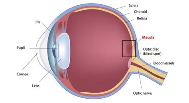
What is macula and fovea?
The macula is the pigmented part of the retina located in the very center of the retina. In the center of the macula is the fovea, perhaps the most important part of the eye. The fovea is the area of best visual acuity. It contains a large amount of cones—nerve cells that are photoreceptors with high acuity.
How does a fovea work?
Fovea: In the eye, a tiny pit located in the macula of the retina that provides the clearest vision of all. Only in the fovea are the layers of the retina spread aside to let light fall directly on the cones, the cells that give the sharpest image. Also called the central fovea or fovea centralis.
What is the function of the ciliary body in the eye?
The ciliary body is found behind the iris and includes the ring-shaped muscle that changes the shape of the lens when the eye focuses. It also makes the clear fluid that fills the space between the cornea and the iris.
Is the fovea responsible for central vision?
The fovea is responsible for sharp central vision (also called foveal vision), which is necessary in humans for activities for which visual detail is of primary importance, such as reading and driving.
What's the difference between Fossa and fovea?
As nouns the difference between fovea and fossa is that fovea is (anatomy) a slight depression or pit in a bone or organ while fossa is (anatomy) a pit, groove, cavity, or depression, of greater or less depth or fossa can be a carnivorous mammal endemic to madagascar,.
What movement is the hip?
The structure of the hip allows a wide range of motion to (and between) the extreme ranges of anterior, posterior, medial, and lateral movement. Raising the leg toward the front is termed flexion; pushing the leg toward the back is termed extension (Figure 2).
What is the lesser trochanter?
The lesser trochanter – A pyramidal prominence that projects from the proximal (near) and medial (inside) part of the shaft of the femur. The lesser trochanter is also called the minor trochanter, the inner trochanter, and the medial process of the femur.
Why would you need a hip replacement?
Hip replacement surgery is usually necessary when the hip joint is worn or damaged so that your mobility is reduced and you are in pain even while resting. The most common reason for hip replacement surgery is osteoarthritis. Other conditions that can cause hip joint damage include: rheumatoid arthritis.
What makes up the obturator foramen?
Anatomical terms of bone The obturator foramen (Latin foramen obturatum) is the large opening created by the ischium and pubis bones of the pelvis through which nerves and blood vessels pass.
What is the danelius Miller method?
The Danelius-Miller method is performed with the detector/image receptor positioned parallel against the affected leg. The primary beam is directed perpendicular to the affected femoral neck region, entering the medial aspect and exiting the lateral aspect of the affected leg.