What are the four independent movements associated with the ankle and the two combination movements at the ankle
The movements that occur at the ankle joint are plantarflexion, dorsiflexion, inversion, and eversion.
Which 2 movements can take place at the ankle joint?
Dorsiflexion and plantar flexion are movements at the ankle joint, which is a hinge joint.
What four bones make up the ankle joint?
- the tibia, the larger and stronger of the two lower leg bones, which forms the inside part of the of the ankle.
- the fibula, the smaller bone of the lower leg, which forms the outside part of the ankle.
- the talus, a small bone between the tibia and fibula and the calcaneus, or heel bone.
What are the main movements that occur at the ankle joint?
The key movement of the ankle joint complex are plantar- and dorsiflexion, occurring in the sagittal plane; ab-/adduction occurring in the transverse plane and inversion-eversion, occurring in the frontal plane8 (Figure 3).What are the two primary movements of the ankle quizlet?
Fundamental ankle joint movements in the sagittal plane include plantar flexion and circumduction. The flexor hallucis longus muscle flexes the big toe and also assists (as a synergist) the gastrocnemius and soleus in plantar flexion of the floor.
What is the function of ankle?
The ankle joint allows up-and-down movement of the foot. The subtalar joint sits below the ankle joint, and allows side-to-side motion of the foot.
What is dorsiflexion and plantar flexion?
The term plantar flexion refers to the movement of the foot in a downward motion away from the body. … It also enables the opposite movement, dorsiflexion, which is the movement of the foot toward the leg. Your ankle joint supplies the power for 40% to 70% of your forward movement during walking.
What bone is the ankle bone?
The talus (or “ankle bone”) connects your leg to your foot.Which blood vessels supply the ankle joint?
Neurovascular Supply The arterial supply to the ankle joint is derived from the malleolar branches of the anterior tibial, posterior tibial and fibular arteries. Innervation is provided by tibial, superficial fibular and deep fibular nerves.
Which three bones make up the ankle joint quizlet?The bones of the ankle include the tibia, fibula, and talus. You just studied 92 terms!
Article first time published onWhat's your ankle bone called?
The hind foot consists of the Talus bone or ankle bone and the calcaneous bone or heel bone. The calcaneous bone is the largest bone in your foot while the talus bone is the highest bone in your foot. The calcaneous joins the Talus bone at the subtalar joint enabling the foot to rotate at the ankle.
Which malleolus is most posterior?
Anatomy of an Ankle Fracture The medial malleolus on the inner side of the ankle at the end of the tibia. The lateral malleolus on the outer side of the ankle at the end of the fibula. The posterior malleolus situated on the lower back side of the tibia.
Which of the 2 malleolus is more distal?
The distal fibula continues to become the lateral malleolus. The lateral malleolus is significantly more evident than the medial malleolus and can be palpated at the ankle.
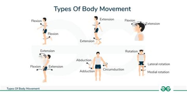
Which type of angular movement moves a body part away from the midline of the body or another reference point?
A movement of a body part away from the midline, either of the body as a whole or that of the hand or foot, is termed abduction (L., to carry away). A movement of the body part back toward the midline (i.e., to the anatomical position) is known as adduction.
What muscles are involved in dorsiflexion and plantar flexion?
There’s one muscle on the front of the leg for dorsiflexion, tibialis anterior. There are three on the back of the leg for plantar flexion, gastrocnemius, soleus, and plantaris. Here’s tibialis anterior.
Which movement is an example of dorsiflexion?
Dorsiflexion is where the toes are brought closer to the shin. This decreases the angle between the dorsum of the foot and the leg. For example, when walking on the heels the ankle is described as being in dorsiflexion.
What are anatomical movements?
Anatomical movements can be defined as the act or instance of moving the bodily structures or as the change of position in one or more of the joints of the body. Joint actions are described in relation to the anatomical position which is the universal starting position for describing movement.
What 4 ligament structures form the ankle syndesmosis?
It is formed between the distal tibia(concave surface) and fibula(convex surface), with no articular capsule or synovial membrane as a fibrous joint, and attached by the interosseous ligament (IOL), the anterior-inferior tibiofibular ligament (AITFL), the posterior-inferior tibiofibular ligament (PITFL), and the …
What muscles are in the ankle?
The major muscles of the ankle include the gastrocnemius and soleus (calf) muscles, which push the foot down and allow us to go up on our toes. These two large muscles join at the ankle to form the Achilles tendon.
Which movement has the greatest range of motion in the ankle joint?
Motion of the ankle occurs primarily in the sagittal plane, with plantar- and dorsiflexion occurring predominantly at the tibiotalar joint. Several studies have indicated an overall ROM in the sagittal plane of between 65 and 75°, moving from 10 to 20° of dorsiflexion through to 40–55° of plantarflexion.
What movements can the ankle do?
In total, the ankle allows the foot to move in six different ways: dorsiflexion, plantarflexion, inversion, eversion, and medial and lateral rotation. Flexion and extension at the ankle are referred to as dorsiflexion and plantarflexion, respectively (Figure 2).
What joint movements occur between the tarsal bones?
Movements. The complex motion of the subtalar joint occurs in three planes and produces subtalar inversion and eversion. Along with the transverse tarsal joint (i.e. talonavicular and calcaneocuboid joint), the subtalar joint transforms tibial rotation into forefoot supination and pronation.
What are the 3 principle arteries that supply the leg and ankle?
The three main arteries which supply the leg and ankle region are all branches of the popliteal artery. They’re the anterior tibial, the posterior tibial, and the peroneal.
What are the ligaments in the ankle?
The major ligaments of the ankle are: the anterior tibiofibular ligament (2), which connects the tibia to the fibula; the lateral collateral ligaments (3), which attach the fibula to the calcaneus and gives the ankle lateral stability; and, on the medial side of the ankle, the deltoid ligaments (4), which connect the …
Why do I have two ankle bones?
It is connected to the talus by a fibrous band. The presence of an os trigonum in one or both feet is congenital (present at birth). It becomes evident during adolescence when one area of the talus does not fuse with the rest of the bone, creating a small extra bone. Only a small number of people have this extra bone.
What happens when you roll an ankle?
A sprained ankle is an injury that occurs when you roll, twist or turn your ankle in an awkward way. This can stretch or tear the tough bands of tissue (ligaments) that help hold your ankle bones together. Ligaments help stabilize joints, preventing excessive movement.
What are the bones in the ankle called quizlet?
Tarsals (the 7 ankle bones)
Which bones are found in the ankle quizlet?
What is the ankle? A hinge joint formed by the articulation of 2 long bones, the tibia, fibula, and the talus.
Which bones make up the foot quizlet?
- Phalanges (bones of foot)
- Metatarsals (bones of instep)
- Tarsals (bones of ankles)
What are toes?
Toes are the digits of the foot. The toe refers to part of the human foot, with five toes present on each human foot. … The first toe, also known as the hallux (“big toe” or “great toe”), the innermost toe. The second toe, or “long toe” The third toe, or “middle toe”
What bones are in wrist?
Your wrist is made up of eight small bones (carpal bones) plus two long bones in your forearm — the radius and the ulna. Each finger consists of one hand bone (metacarpal) and three finger bones (phalanges), while each thumb consists of one metacarpal bone and two phalanges.