What are Kerley A lines
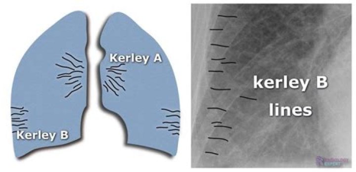
Kerley A lines are linear opacities extending from the periphery to the hila caused by distention of anastomotic channels between peripheral and central lymphatics. Kerley B lines are small, horizontal, peripheral straight lines demonstrated at the lung bases that represent thickened interlobular septa on CXR.
What do Kerley B lines represent?
Kerley B lines (arrows) are horizontal lines in the lung periphery that extend to the pleural surface. They denote thickened, edematous interlobular septa often due to pulmonary edema.
What are B lines on ultrasound?
B lines, previously termed ”comet tails,” are vertical hyperechoic reverberations moving synchronously with the lung and represent key artifacts in interpreting pulmonary ultrasound findings [3,4]. The physiologic basis of B lines relates to decreased lung aeration [5], a finding that is nonspecific.
Are Kerley lines normal?
Kerley A lines On chest radiographs they are seen to cross normal vascular markings and extend radially from the hilum to the upper lobes.How would you describe pulmonary edema on CXR?
At chest radiography and CT, postobstructive pulmonary edema typically manifests as septal lines, peribronchial cuffing, and, in more severe cases, central alveolar edema (,Fig 10). These findings are similar to those in pressure edema.
What swollen lungs?
Pulmonary edema is a condition caused by excess fluid in the lungs. This fluid collects in the numerous air sacs in the lungs, making it difficult to breathe. In most cases, heart problems cause pulmonary edema.
What is Hampton hump?
Hampton’s hump is a radiological sign consisting of a peripheral, wedge-shaped opacification adjacent to the pleural surface, which represents pulmonary infarction distal to a pulmonary embolus. 1. Owing to good pulmonary perfusion from collateral blood vessels, this sign is rarely seen in clinical practice.
What is alveolar edema?
Pulmonary edema occurs when fluid accumulates in the air sacs of the lungs – the alveoli – making it difficult to breathe. This interferes with gas exchange and can cause respiratory failure. Pulmonary edema can be acute (sudden onset) or chronic (occurring more slowly over time).What is a phantom tumor?
— The “phantom tumor” is a roentgenographic finding caused by fluid localizing in an interlobar fissure of the lung secondary to congestive heart failure. These fluid accumulations have also been called “vanishing tumor” and “pseudo tumor” of the lung because they tend to disappear when the heart failure is treated.
What Orthopnea means?Orthopnea is the sensation of breathlessness in the recumbent position, relieved by sitting or standing. Paroxysmal nocturnal dyspnea (PND) is a sensation of shortness of breath that awakens the patient, often after 1 or 2 hours of sleep, and is usually relieved in the upright position.
Article first time published onWhat do B lines mean?
The B-line is a kind of comet-tail artifact indicating subpleural interstitial edema. The relationship between anterior interstitial edema detected by lung ultrasound and the pulmonary artery occlusion pressure (PAOP) value was investigated.
What do B lines represent?
B lines indicate filling of intralobular or interlobular septa and are often seen in pulmonary edema and interstitial lung diseases. Thickened B lines may fuse together to form coalescent B lines representing peripheral lung ground glass opacities seen in high resolution computed tomography (CT).
Where can I find B lines?
Experts recommend scanning at least 1 posterior zone (posterior to posterior axillary line) on each side as well in supine patients when monitoring response to diuretic or ultrafiltration therapy. Visualization of occasional B-lines, especially in the dependent zones of the lung is not abnormal.
How can you tell the difference between pulmonary edema and pneumonia?
The major difference being that pneumonia is an infectious pathology while pulmonary edema is not usually caused by an infection. It is a marker for a more severe underlying systemic pathology like heart failure or volume overload states in the body.
Do steroids help pulmonary edema?
High-dose prednisolone reduced EVLW and improved hemodynamics and gas exchange in patients with noncardiac pulmonary edema, whereas placebo did not achieve comparable effects. Therefore, high-dose prednisolone appears beneficial in noncardiac pulmonary edema in respect of EVLW, hemodynamics, and gas exchange.
What does Cephalization mean in radiology?
Cephalization refers to the redistribution of blood into the upper lobe vessels. It has been hypothesized that once the hydrostatic pressure exceeds 10 mm Hg, then fluid begins to leak into the interstitium of the lung. This excess fluid initially compresses the lower lobe vessels, perhaps as a result of gravity.
What is S1Q3T3?
However, the “S1Q3T3” pattern of acute cor pulmonale is classic; this is termed the McGinn-White Sign. Enlarge. A large S wave in lead I, a Q wave in lead III and an inverted T wave in lead III together indicate acute right heart strain.
What is consolidation in the lung?
Lung consolidation occurs when the air that usually fills the small airways in your lungs is replaced with something else. Depending on the cause, the air may be replaced with: a fluid, such as pus, blood, or water. a solid, such as stomach contents or cells.
What is pulmonary plethora?
Plethoric lung fields are seen in conditions which increase pulmonary blood flow.
What do damaged lungs feel like?
Coughing up blood: If you are coughing up blood, it may be coming from your lungs or upper respiratory tract. Wherever it’s coming from, it signals a health problem. Chronic chest pain: Unexplained chest pain that lasts for a month or more—especially if it gets worse when you breathe in or cough—also is a warning sign.
Can lungs hurt in your back?
Pleurisy, which is inflammation in the lining of the lungs, can cause sharp pains in the back and chest. This can often be the result of a viral or bacterial infection. Asthma, a chronic, long-term infection of the lung, may also cause pain in your back. Costochondritis is inflammation of rib cage cartilage.
How long does Covid pneumonia last?
For the 15% of infected individuals who develop moderate to severe COVID-19 and are admitted to the hospital for a few days and require oxygen, the average recovery time ranges between three to six weeks.
What is a Hydropneumothorax?
Hydropneumothorax is the abnormal presence of air and fluid in the pleural space. The knowledge of hydropneumothorax dates back to the days of ancient Greece when the Hippocratic succussion used to be performed for the diagnosis.
What is a pseudotumor in the brain?
Pseudotumor cerebri (SOO-doe-too-mur SER-uh-bry) occurs when the pressure inside your skull (intracranial pressure) increases for no obvious reason. It’s also called idiopathic intracranial hypertension. Symptoms mimic those of a brain tumor.
What is LLL pleural effusion?
Pleural effusion, sometimes referred to as “water on the lungs,” is the build-up of excess fluid between the layers of the pleura outside the lungs. The pleura are thin membranes that line the lungs and the inside of the chest cavity and act to lubricate and facilitate breathing.
How can I remove fluid from my lungs at home?
- Steam therapy. Steam therapy, or steam inhalation, involves inhaling water vapor to open the airways and help the lungs drain mucus. …
- Controlled coughing. …
- Drain mucus from the lungs. …
- Exercise. …
- Green tea. …
- Anti-inflammatory foods. …
- Chest percussion.
What does swollen feet and shortness of breath mean?
Congestive heart failure. As a result, blood can back up in your legs, ankles and feet, causing edema. Congestive heart failure can also cause swelling in your abdomen. Sometimes, this condition can cause fluid to accumulate in your lungs (pulmonary edema), which can lead to shortness of breath.
How do you drain fluid from your lungs?
Thoracentesis is a procedure in which a needle is inserted into the pleural space between the lungs and the chest wall. This procedure is done to remove excess fluid, known as a pleural effusion, from the pleural space to help you breathe easier.
What causes nocturnal dyspnea?
PND is caused by the failure of the left ventricle. When this happens, it is unable to pump as much blood as the right ventricle, which is functioning normally. As a result, you experience pulmonary congestion, a condition in which fluid fills the lungs.
Why is it harder to breathe at night?
your sleeping position puts pressure on your diaphragm. mucus builds up in your throat causing you to cough and struggle for breath. your hormones change at night. your sleeping environment triggers your asthma.
What's the difference between dyspnea and orthopnea?
The medical term for shortness of breath is dyspnea. Orthopnea is a type of dyspnea that only occurs when a person is lying down. People often describe orthopnea as a sensation of tightness in the chest that makes breathing difficult or uncomfortable. Some individuals may also experience chest pain.