Is the maxillary sinus a bone
The maxillary sinus (or antrum of Highmore) is a paired pyramid-shaped paranasal sinus within the maxillary bone which drains via the maxillary ostium into the infundibulum, then through hiatus semilunaris into the middle meatus
Is sinus a bone?
The sinuses are hollow spaces in the skull and the face bones around your nose. There are four pairs of sinuses, named for the bones that they’re located in: The maxillary sinuses are located on each side of your nose, near the cheek bones. The frontal sinuses are located above the eyes, near your forehead.
What bone is the maxilla?
The maxilla is the bone that forms your upper jaw. The right and left halves of the maxilla are irregularly shaped bones that fuse together in the middle of the skull, below the nose, in an area known as the intermaxillary suture. The maxilla is a major bone of the face.
What bones make up the maxillary sinus?
Found in the body of the maxilla, this sinus has three recesses: an alveolar recess pointed inferiorly, bounded by the alveolar process of the maxilla; a zygomatic recess pointed laterally, bounded by the zygomatic bone; and an infraorbital recess pointed superiorly, bounded by the inferior orbital surface of the …What is a maxillary sinus?
(MAK-sih-LAYR-ee SY-nus) A type of paranasal sinus (a hollow space in the bones around the nose). There are two large maxillary sinuses, one in each of the maxillary bones, which are in the cheek area next to the nose. The maxillary sinuses are lined with cells that make mucus to keep the nose from drying out.
Is maxilla same as maxillary bone?
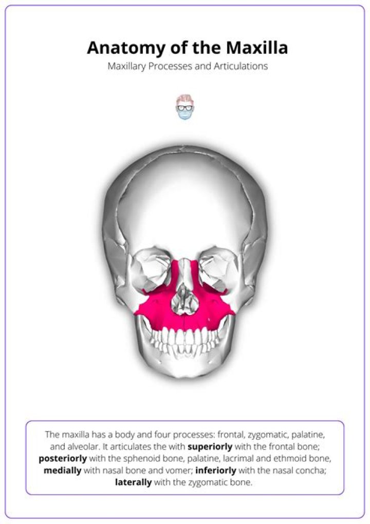
The two maxilla or maxillary bones (maxillae, plural) form the upper jaw (L., mala, jaw). Each maxilla has four processes (frontal, zygomatic, alveolar, and palatine) and helps form the orbit, roof of the mouth, and the lateral walls of the nasal cavity.
Which bones are examples of cranial bones?
- Parietal (2)
- Temporal (2)
- Frontal (1)
- Occipital (1)
- Ethmoid (1)
- Sphenoid (1)
Is maxillary sinus part of maxilla?
The maxillary sinus, or antrum of Highmore, lies within the body of the maxillary bone and is the largest and first to develop of the paranasal sinuses. The alveolar process of the maxilla supports the dentition and forms the inferior boundary of the sinus.What are features of the maxillary bone?
Each maxillary bone has the shape of a pyramid, it’s base adjacent to the nasal cavity, its apex being the zygomatic process, and its body constituting the maxillary sinus. [3] The maxilla connects with surrounding facial structures through four processes: alveolar, frontal, zygomatic and palatine.
What is a maxillary sinus fracture?Maxillary sinus fractures (MSFs) are most commonly caused by blunt force trauma to the face. Depending on the magnitude and location of the direct injury, MSFs can vary in appearance and symptomatology.
Article first time published onWhat is the inferior maxillary bone?
Inferior: nasal concha. Laterally: zygomatic bone. Body of the maxilla. It contains the maxillary sinuses and contributes to the floor of the orbit, lateral wall of the nasal cavity, and anterior part of the infratemporal fossa.
What is right maxillary sinus?
The maxillary sinus is one of the four paranasal sinuses, which are sinuses located near the nose. The maxillary sinus is the largest of the paranasal sinuses. The two maxillary sinuses are located below the cheeks, above the teeth and on the sides of the nose.
Where is vomer bone?
The vomer is a small, thin, plow-shaped, midline bone that occupies and divides the nasal cavity. It articulates inferiorly on the midline with the maxillae and the palatines, superiorly with the sphenoid via its wings, and anterosuperiorly with the ethmoid.
Where is the maxillary ostium?
The first, the maxillary ostium, is located on the superomedial aspect of the maxillary sinus. The ostium leads into the second passage, the ethmoid infundibulum, that conducts mucus from the maxillary sinus into the middle meatus via the third passage, the hiatus semilunaris.
What bones form the cheek?
The zygomatic bone forms the bony prominence of the cheek. It also forms the lower lateral part of the orbital margin, and this part of the lateral orbital wall. The zygomatic bone extends backward to meet the zygomatic process of the temporal bone, forming the zygomatic arch.
Which of the following bones do not have sinuses?
The temporal bone does not contain a sinus.
Which bones contain sinuses?
Paranasal sinuses are named after the bones that contain them: frontal (the lower forehead), maxillary (cheekbones), ethmoid (beside the upper nose), and sphenoid (behind the nose).
What is the posterior wall of the maxillary sinus?
The pterygopalatine fossa, also known as the sphenopalatine fossa, can be thought of as a box, with its anterior wall being the posterior wall of the maxillary sinus. The posterior wall is the pterygoid plates and inferior aspect of the sphenoid bone, the roof is the inferior orbital fissure.
Which of the following bones is not a facial bone quizlet?
True. ETHMOID BONE is one of eight CRANIAL BONES, not the facial bone!
Is the maxillary sinus sterile?
The sinuses are normally sterile under physiologic conditions. Secretions produced in the sinuses flow by ciliary action through the ostia and drain into the nasal cavity.
How do you fix a maxillary sinus fracture?
In the surgical treatment of an isolated anterior wall of the maxillary sinus fracture, open reduction and ridged fixation is required. Large fragments can be fixed with plates and screws and small fragments can either be positioned at the bony defect or removed.
How is a maxillary fracture treated?
Maxillary fractures are treated by reduc- tion and immobilization. Establishment of preinjury occlusion and midface buttress alignment provides the foundation for this treatment. The goals of treatment of LeFort fractures are to reestablish preinjury occlusion with normal height and projection of the face.
Can you damage maxillary sinus?
Paranasal sinuses are air-filled cavities surrounding the nasal cavity proper which includes maxillary sinus, sphenoid sinus, frontal sinus and ethmoid sinus. Trauma to the superior and middle thirds of the face can often lead to in paranasal sinus fractures involving one or more paranasal sinuses.
Where is the maxillary bone located?
The maxilla is a bone which helps to make up the skull. It is specifically located in the mid face, forms the upper jaw, separates the nasal and oral cavities, and contains the maxillary sinuses (located on each side of the nose.
Why do we need sinuses in your facial bones?
The sinuses lighten the skull or improve our voices, but their main function is to produce a mucus that moisturizes the inside of the nose. This mucus layer protects the nose from pollutants, micro-organisms, dust and dirt.
Why do we need sinuses in your facial bones describe why fluid in the maxillary sinus or a sinus infection cause teeth to ache?
When the sinuses become blocked, trapped germs can lead to infection. Once infected, the blocked sinuses may swell and cause pressure in the face. A sinus infection can cause toothache because the swelling and build-up of mucus inside the sinuses may put pressure on nerves running to the roots of the teeth.
How do you unblock a maxillary sinus?
- Place each of your index and middle fingers on either side of your nose, just between your cheekbones and upper jaw. Try using your thumbs instead of your index fingers for stronger pressure.
- Gently massage this area using a circular motion.
- Repeat for around 30 seconds to a minute.
Is vomer a cranial bone?
Located in the center of the nasal cavity, the vomer is a thin, unpaired bone of the face and skull (cranium).
What type of bone is the vomer?
1. Flat Bones Protect Internal Organs. There are flat bones in the skull (occipital, parietal, frontal, nasal, lacrimal, and vomer), the thoracic cage (sternum and ribs), and the pelvis (ilium, ischium, and pubis). The function of flat bones is to protect internal organs such as the brain, heart, and pelvic organs.
What are Conchae?
The conchae are structures made of bone inside of your nose. They help control the airflow into your nose. They also clean and warm air that you’ve inhaled so that it’s ready to go to your lungs for respiration. Respiration is the process of breathing in and out.
Which bones reduce the size of maxillary hiatus?
In the articulated skull this aperture is much reduced in size by the following bones: the uncinate process of the ethmoid above, the ethmoidal process of the inferior nasal concha below, the vertical part of the palatine behind, and a small part of the lacrimal above and in front; the sinus communicates with the …