How do you pull a femoral sheath
Take your index, middle and sometimes your ring finger, and place them slightly above the sheath to feel the patient’s pulse. … Slowly remove the sheath in a sterile manner, holding occlusive pressure to avoid bleeding.
What should a act be before pulling a sheath?
Before removing the sheath, check that the heparin is stopped, the activated clotting time (ACT) is less than 150 seconds, vital signs are stable, no chest pain is present, and there are no plans for recatheterization.
How do you pull a FEM line?
Cleanse site with 2% chlorhexidine and 70% alcohol swab and remove any sutures. Gently withdraw catheter while applying direct pressure with the sterile gauze. Stop withdrawal and notify physician if the catheter does not withdraw easily. Hold pressure until physician assesses limb if partial withdrawal occurs.
When do you remove femoral sheath?
The anticoagulation time (ACT) should ideally be less than 160 seconds (Grossman and Baim, 2000). In practice, it is time consuming trying to measure the ACT. Therefore it is our local practice to remove femoral sheaths four hours after the procedure unless the cardiologist specifies otherwise.How do you hold pressure on your femoral artery?
Firm three-finger pressure should control most femoral bleeding. A rolled gauze pack may be placed over the artery to the groin, and pressure applied with the palm of the hand. Standing on a short stool at bedside permits the operator’s upper body weight to be used for pressure application.
What is sheath size?
Sheath sizes range from 4 French (Fr) to 24 Fr for percutaneous procedures, with most using 4-6 Fr for diagnostic angiography. Sheath sizes that exceed 10 Fr are usually reserved for special procedures, with the largest used for procedures such as transcatheter valve replacement.
How do you pull a groin sheath?
- Take your index, middle and sometimes your ring finger, and place them slightly above the sheath to feel the patient’s pulse. …
- Slowly remove the sheath in a sterile manner, holding occlusive pressure to avoid bleeding.
What is femoral catheterization?
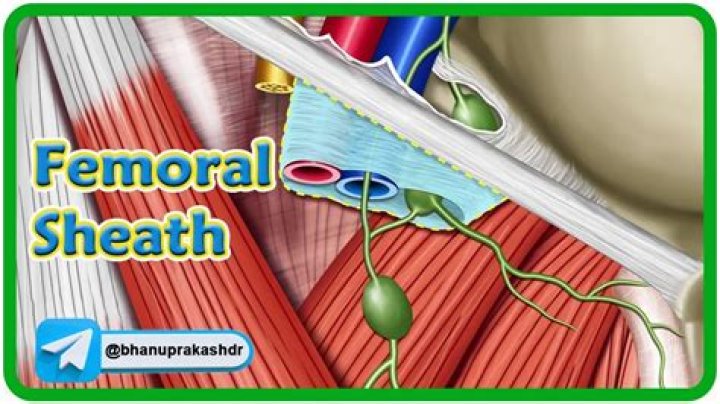
Percutaneous cannulation of the femoral vein uses anatomic landmarks to guide venipuncture and a Seldinger technique to thread a central venous catheter through the femoral vein and into the inferior vena cava.What is a femoral sheath?
The femoral sheath is a structure within bilateral femoral triangles. The femoral sheath contains the femoral vein, artery, and lymphatics. The femoral nerve lies lateral to the femoral sheath and is not enclosed within the sheath.[1][2]
How do you remove a femoral PICC line?- Apply Related Procedures and Policies.
- Check Coagulation Tests.
- Prepare Bedside.
- Prepare Tray.
- Remove Dressing.
- Cleanse Site and Remove Suture.
- Remove Catheter.
- Ensure Hemostasis.
How do you insert a femoral line?
The site to choose should be 1-2 cm below the inguinal crease, about 1cm medial to the femoral pulse. Be sure to enter below the inguinal crease to avoid retroperitoneal puncture. Using the blue 25 ga needle anesthetize the skin with lidocaine, and then the subcutaneous tissues with the green 22 ga needle.
Is central line removal painful?
It can become painful to be repeatedly poked with needles or fitted with IVs. To help limit your discomfort during treatments, a long-term IV or central line may be an option.
Where do you get a femoral puncture?
The ideal site of femoral arterial puncture (not skin puncture) is at the CFA at a point approximately 1 cm lateral to the most medial aspect of the femoral head, midway between its superior and inferior borders (Rupp’s rule).
Which leg is the femoral artery in?
The femoral artery is the major blood vessel supplying blood to your legs. It’s in your upper thigh, right near your groin.
How do you close the femoral artery?
Traditionally, the main method of closing the artery is compression, during which up to 30 minutes of manual pressure or mechanical clamps is applied directly to the patient’s groin. This manual pressure can be painful and requires up to eight hours of bedrest.
What is a radial sheath?
The radial artery access sheath is used to gain arterial access and facilitate the insertion of catheters or other equipment for diagnostic and vascular interventions.
What is a sheath cardiac?
During a cardiac catheterization, a long, narrow tube called a catheter is inserted through a plastic introducer sheath (a short, hollow tube that is inserted into a blood vessel in your leg or arm).
What is an angioplasty?
A coronary angioplasty is a procedure used to widen blocked or narrowed coronary arteries (the main blood vessels supplying the heart). The term “angioplasty” means using a balloon to stretch open a narrowed or blocked artery.
Is sheath a catheter?
Penile Sheath Sheaths are a popular and effective alternative to absorbent pads for many men. … Penile Sheaths are also sometimes called condom catheters, uridomes or external catheters.
How does introducer sheath work?
An introducer set contains devices used to access blood vessels for the insertion of vascular catheters. After a needle is inserted through the skin and into the blood vessel, the dilator and sheath are passed together into the blood vessel.
What is a introducer sheath used for?
Introducer sheaths are used when there is the need for a larger catheter, such as for rapid volume administration or for the insertion of a pulmonary artery catheter.
What lies in the femoral sheath?
The femoral sheath is contained in the femoral triangle. … The lateral compartment contains the femoral artery, and the femoral branch of genitofemoral nerve. The intermediate compartment contains the femoral vein.
Why femoral nerve is outside the sheath?
The femoral nerve is the largest branch of the lumbar plexus. … The nerve enters the femoral triangle by passing beneath the inguinal ligament, just lateral to the femoral artery. In the thigh, it lies outside the femoral sheath, gives off articular branches to the hip and knee joints.
What goes through the femoral canal?
- Lymphatic vessels – draining the deep inguinal lymph nodes.
- Deep lymph node – the lacunar node.
- Empty space.
- Loose connective tissue.
Why is femoral catheterization done?
The following are generally accepted indications for femoral venous catheter placement: Emergency venous access during cardiopulmonary resuscitation (CPR), in that it provides a rapid and reliable route for the administration of drugs to the central circulation of the patient in cardiac arrest.
Can you walk with a femoral line?
Physical therapy sessions, including standing and walking were feasible and safe in cardiovascular ICU patients with femoral catheters who met the criteria for mobility interventions.
Is femoral vein superficial or deep?
In particular, the femoral vein is clinically a deep vein, where deep vein thrombosis indicates anticoagulant or thrombolytic therapy, but the adjective “superficial” leads many physicians to falsely believe it is a superficial vein, which has resulted in patients with femoral thrombosis being denied proper treatment.
Can you draw labs from an arterial line?
Blood drawing from indwelling arterial or central venous lines is done through a stopcock with a needleless access device on the sampling port.
Who can remove arterial line?
4. Radial artery arterial lines can be removed by an RN deemed competent in the procedure. 1. The purpose of an arterial line is to identify changes in arterial pressure, evaluate medical therapy (i.e. titration of drugs, fluid administration) and obtain blood samples.
How do you pull a PICC line?
Hold the sterile gauze in one hand (ready to put it over the insertion site when the catheter comes out) and with the other hand grasp the hub and main catheter. Gently and steadily pull out the catheter, moving your hand closer to the insertion site as you remove the PICC. Stop pulling if you feel resistance.
How do you remove a perm from a catheter?
Doctor makes a small incision and removes the catheter from the vein. In this, doctor uses two hemostats to release the cuff from the tissue poking deep into the hole by helding pressure at the jugular (entrance site under the skin) and slides the catheter out.